Session Information
Date: Saturday, November 6, 2021
Title: Systemic Sclerosis & Related Disorders – Clinical Poster I (0387–0413)
Session Type: Poster Session A
Session Time: 8:30AM-10:30AM
Background/Purpose: There are currently no validated objective measures of systemic sclerosis (SSc)-related skin thickening and this hampers clinical trials of potential new treatments. The modified Rodnan skin score (mRSS, the current gold standard) has limitations including concerns about inter-rater reliability. The aim of this study was to test the hypothesis that high frequency ultrasound (HFUS), which measures skin thickness is (a) sensitive to change and (b) correlates with local ‘skin score’ (criterion validity), both OMERACT criteria for validation.
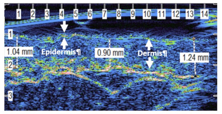
Methods: In this prospective study, patients with early diffuse cutaneous SSc (dcSSc; duration of first non-Raynaud’s clinical manifestation < 5 years) underwent HFUS at each visit. Imaging was carried out at nine sites (most corresponding to sites assessed for mRSS; non-dominant ring finger, dorsum of hand, forearm, upper arm, forehead, anterior chest, abdomen, leg and dorsum of foot. Local skin score (0-3; 0 normal, 3 unable to move) was also assessed at each site. Images were assessed by one observer; skin thickness was measured as the distance from the surface of the skin to the base of the dermis (i.e. epidermis and dermis). The average thickness was taken of 3 measures made on each image at the left, centre and right in order to take into account any differences in thickness across the image (Figure 1). Longitudinal data underwent linear mixed effects modelling; a random intercept model with a Holm- Bonferroni correction applied.
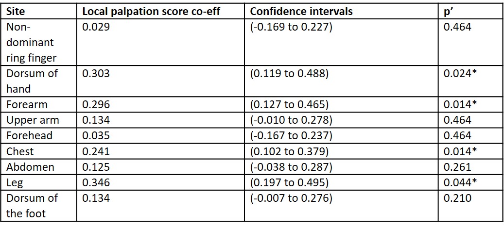
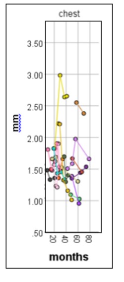
Results: Seventeen patients with dcSSc were recruited (10 F:7M), median age 54 (range 42 to 61) years, median duration of Raynaud’s 2 (IQR 2- 5) years and duration of first non-Raynaud’s clinical manifestation 2 (1- 4) years. Three patients had 5 sets of measures, four had 4, four had 3, six had 2; median duration of follow-up of 16 months (range 7-43 months). Although patients did not follow a set pattern of change (i.e. skin thickness increased for some patients and sites and decreased for others) HFUS was sensitive to changes in thickness (example in Figure 2). Modelling indicated that changes in local skin score tracked well with changes in HFUS thickness measures at the dorsum of the hand, forearm, chest and leg (indicated by the local skin score coefficent in Table 1, p’< 0.05).
Conclusion: Conclusions
In patients with early dcSSc HFUS was able to determine differences in skin thickness with time, indicative of sensitivity to change.
Changes in skin thickness as measured by HFUS mirrored changes assessed by local palpation scores at several sites.
Patients at first visit had different levels of skin thickening due to the different durations and
severities of disease, this heterogeneity makes identifying patterns of change over time in patients difficult.
These findings support the hypothesis that HFUS is sensitive to change and shows criterion validity, showing promise for future validation of HFUS as an outcome measure in clinical trials.
To cite this abstract in AMA style:
Marjanovic E, Heal C, Moore T, Manning J, Wilkinson S, Dinsdale G, Dickinson M, Wilkinson J, Herrick A, Murray A. High Frequency Ultrasound Imaging of Skin Thickness in Patients with Early Diffuse Cutaneous Systemic Sclerosis: An Objective Outcome Measure to Track Change over Time [abstract]. Arthritis Rheumatol. 2021; 73 (suppl 9). https://acrabstracts.org/abstract/high-frequency-ultrasound-imaging-of-skin-thickness-in-patients-with-early-diffuse-cutaneous-systemic-sclerosis-an-objective-outcome-measure-to-track-change-over-time/. Accessed .« Back to ACR Convergence 2021
ACR Meeting Abstracts - https://acrabstracts.org/abstract/high-frequency-ultrasound-imaging-of-skin-thickness-in-patients-with-early-diffuse-cutaneous-systemic-sclerosis-an-objective-outcome-measure-to-track-change-over-time/