Session Information
Date: Tuesday, November 14, 2023
Title: (2352–2369) Systemic Sclerosis & Related Disorders – Clinical Poster III: Translational Science
Session Type: Poster Session C
Session Time: 9:00AM-11:00AM
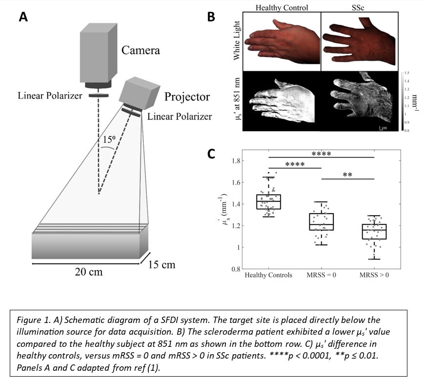
Background/Purpose: Systemic sclerosis (SSc) is an autoimmune disease characterized by excessive collagen deposition in the skin and internal organs, along with vascular dysfunction. The modified Rodnan skin score (mRSS) is the established gold standard for evaluating skin fibrosis in SSc. Nevertheless, mRSS is not objective, has significant interrater variability and may miss small but clinically significant changes. Spatial Frequency Domain Imaging (SFDI) is a non-invasive and optical technique that uses near-infrared light to generate widefield images of tissue optical properties (absorption (μa) and reduced scattering coefficients (μs‘)) at sub-surface depths. Our recent study showed that μs´ is highly correlated with mRSS, suggesting that SFDI is a reliable method for assessing skin fibrosis in SSc patients (Fig. 1) (1). However, the sensitivity of SFDI to longitudinal changes has yet to be examined.
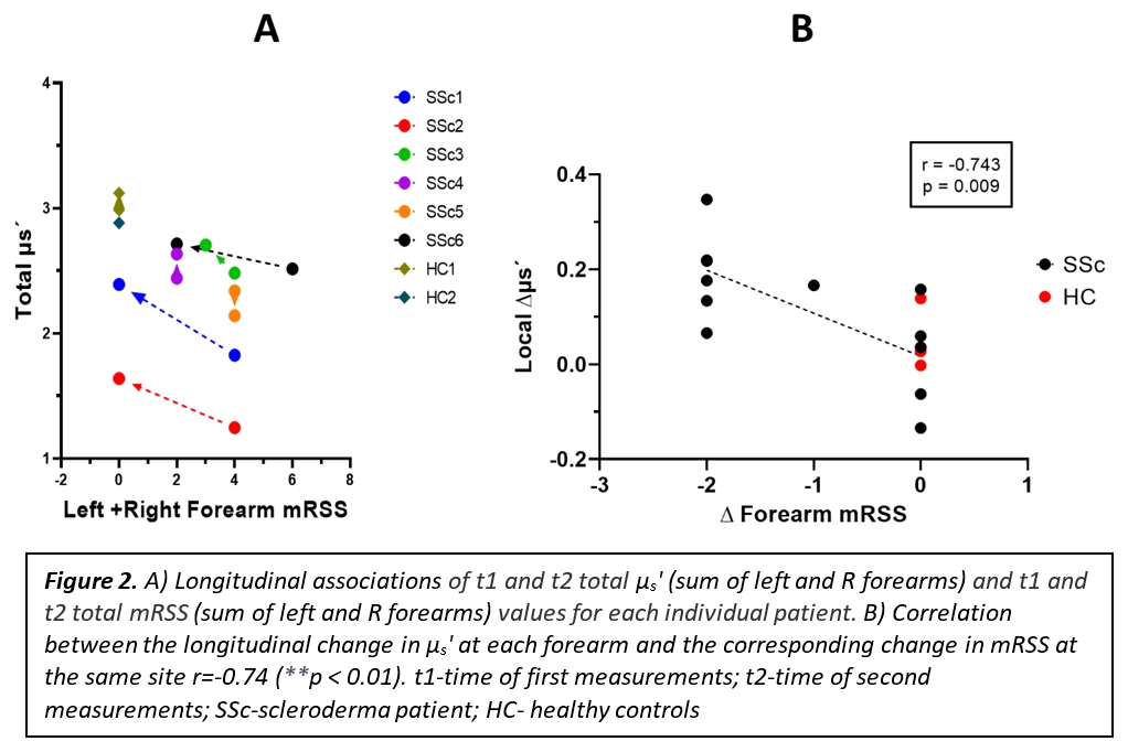
Methods: The SFDI data were collected from the left and right forearms in 2 healthy control (HC) subjects and 6 patients with diffuse SSc.Subjects were measured at two different time points, at least two months apart. Local forearm mRSS scores were performed by a trained physician, blinded to the SFDI measurements and using the averaging method. A rectangular region of interest (ROI) of dimensions 800 × 300 pixels (approx. 4.3 × 11.5 cm), aligned along the dorsal forearm was used to measure μs‘ at 851 nm .Differences between the sum of left and right forearms in each patient were plotted against differences in the sum ofμs‘ in each patient. Correlation between the absolute change of μs‘ versus absolute change of mRSS at the 2 time-points were evaluated using the Spearman test. A p-value < 0.05 was considered statistically significant. All data were analyzed with GraphPad Prism 9 software.
Results: 83% of SSc patients were females, with a mean age of 44 ± 18.4 years. Mean disease duration was 4.3 ± 2.6 years and mean follow up interval between measurements was 23 months ± 4.8 months. In 3 patients the sum of forearm mRSS scores improved by 4 points (SSc1, 2 and 6), one patient had a modest improvement (1 point, SSc3), while two patients remained stable (SSc4 and 5, Fig 2A). No patient had an increase in the forearm skin score. As expected, the patients with no improvement showed minimal changes in the μs’, similar to controls (0.0109 ± 0.1129 mm-1 in SSc compared to 0.0497 ± 0.0613 mm-1 in HC). However, when patients improved there was a proportionate increase in μs’ that mirrored the degree of mRSS improvement (Fig 2A). Next we assessed correlation between change in mRSS and change in μs’ at each site, and found a significant negative correlation (r = -0.74, p < 0.01, Fig 2B).
Conclusion: Our preliminary study indicates that SFDI is sensitive to longitudinal changes in the skin of SSc patients, further supporting this method as a highly promising new tool for scleroderma skin assessment and monitoring. Future studies are required to confirm these findings.
References:
1. Pilvar A, Mehendale AM, Karrobi K, El-Adili F, Bujor A, Roblyer D. Spatial frequency domain imaging for the assessment of scleroderma skin involvement. Biomed Opt Express. 2023;14(6):2955. doi:10.1364/BOE.489609
To cite this abstract in AMA style:
Vo H, Mehendale A, Pilvar A, Kissin E, Trojanowski M, York M, Roblyer D, Bujor A. Spatial Frequency Domain Imaging as a Novel Method to Quantify Longitudinal Skin Changes in Scleroderma [abstract]. Arthritis Rheumatol. 2023; 75 (suppl 9). https://acrabstracts.org/abstract/spatial-frequency-domain-imaging-as-a-novel-method-to-quantify-longitudinal-skin-changes-in-scleroderma/. Accessed .« Back to ACR Convergence 2023
ACR Meeting Abstracts - https://acrabstracts.org/abstract/spatial-frequency-domain-imaging-as-a-novel-method-to-quantify-longitudinal-skin-changes-in-scleroderma/