Session Information
Date: Tuesday, October 28, 2025
Title: (1780–1808) Osteoarthritis & Joint Biology – Basic Science Poster
Session Type: Poster Session C
Session Time: 10:30AM-12:30PM
Background/Purpose: Osteoarthritis (OA) is a joint pathology involving joint cartilage destruction and synovial inflammation. Severe knee OA is often surgically treated with total knee arthroplasty (TKA) joint replacement. Approximately 20% of patients experience continued pain and dysfunction postoperatively, but no predictors of outcome are known. We previously reported that bacterial peptidoglycan (PG) is associated with increased synovitis and elevated IL-6. However, PG did not correlate with patient reported outcomes (PRO) post-TKA, indicating other immune mediators may be contributing to surgical outcome. We therefore used highly multiplexed cell imaging to identify cell-cell interactions associated with incomplete responses to surgery and other disease features.
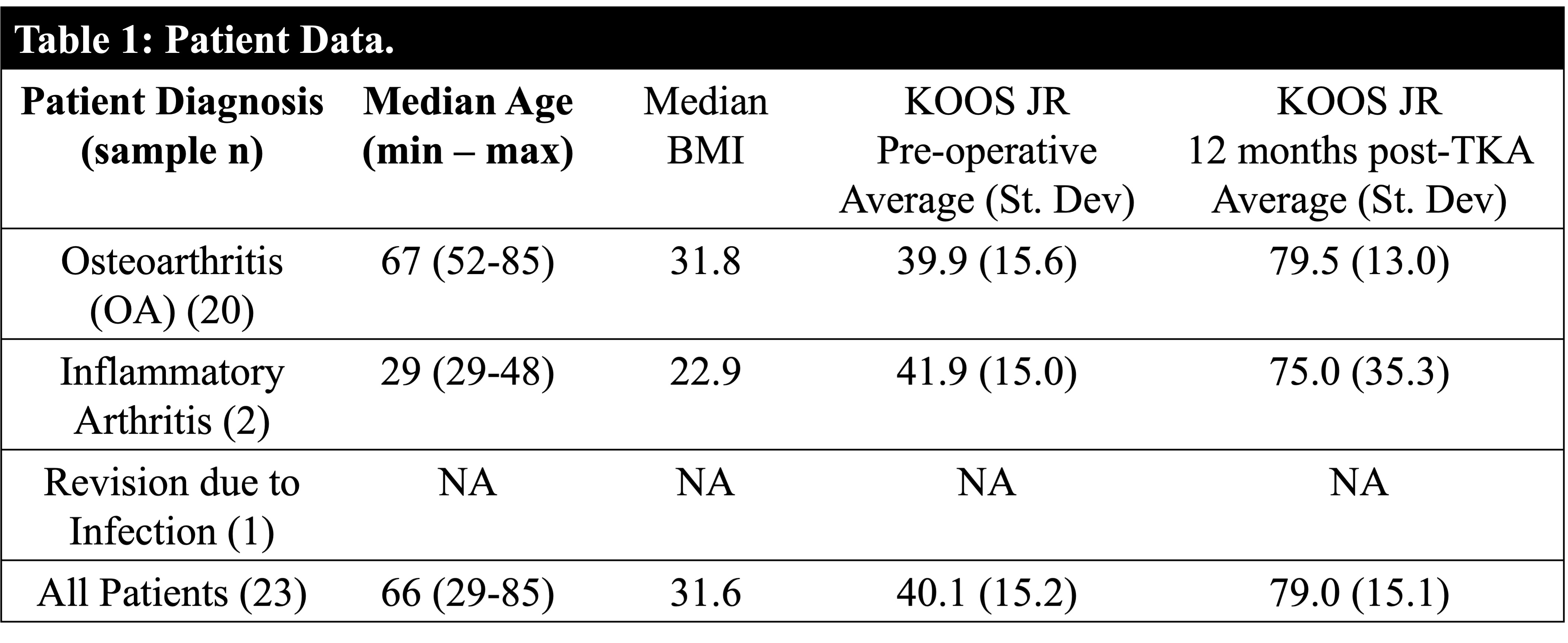
Methods: Discarded synovial tissue (ST) were collected from 23 patients undergoing primary TKA, 20 (87%) of which met ACR criteria for knee OA (Table 1). Knee Osteoarthritis Outcome Score – Joint Replacement (KOOS JR) PRO were collected at pre-operative and 3-, 6-, and 12-month post-operative time points. Multiplexed immunofluorescence microscopy (Akoya Biosciences) was used to identify T cells (CD3, CD4, CD8), antigen presenting cells (HLA-DR, CD11c), endothelial cells (CD31), plasma cells (CD138), and stromal cells (CD90, CD34) in synovial tissue. Cell-cell distance was calculated to infer cell interaction using the Scimap Python package. For the patients with advanced knee OA, associations between spatial imaging data and disease features, including PRO scores, were determined by calculating Pearson’s r correlation coefficients using Prism software (v.10).
Results: Patient synovitis was highly variable, consisting of infiltrate-rich inflammatory foci and/or regions of vascular inflammation and fibrosis (Fig 1A). Vascular lesions typically contained CD4+ T cells near CD31+ endothelial cells (Fig 1B). Inflammatory foci were enriched in both CD4+ T cells and CD8+ T cells (Fig 1C). Total CD4+ T cells correlated with PG in tissue, whereas total CD8+ T cells correlated with overall synovial lesion score (Fig 1D). CD4-CD31 spatial distance tended to be shortest in patients with low/variable PRO score 12 months post-TKA (Pearson’s r=0.512, P=0.074), whereas CD8-CD4 spatial distance was associated with inflammatory synovitis.
Conclusion: Spatial imaging of advanced OA ST revealed an important role in T cells in contributing to synovitis. In our cohort, average distances between CD4+ T-helper cells and CD31+ endothelial cells in ST at time of surgery correlated with low 12-month PRO score, suggesting that vascular inflammation in OA synovitis may contribute to poor or variable long-term outcome post-TKA.
.jpg) Figure 1. Spatial imaging analysis of advanced OA synovial tissue. (A) Representative images are shown of OA synovial tissue is shown using cell markers indicated in figure; (B) a representative image of CD4 T cells in close proximity to CD31+ endothelial cells; (C) and a representative image of typical inflammatory foci containing CD4 and CD8 T cells. (D) Associations between total T cell numbers and cell-cell spatial distances with disease features. Pearson’s R and P values are indicated in figure. For spatial distance analyses, only OA synovial tissue were included, and spatial distance is shown in pixels (1 pixel≅2 μm).
Figure 1. Spatial imaging analysis of advanced OA synovial tissue. (A) Representative images are shown of OA synovial tissue is shown using cell markers indicated in figure; (B) a representative image of CD4 T cells in close proximity to CD31+ endothelial cells; (C) and a representative image of typical inflammatory foci containing CD4 and CD8 T cells. (D) Associations between total T cell numbers and cell-cell spatial distances with disease features. Pearson’s R and P values are indicated in figure. For spatial distance analyses, only OA synovial tissue were included, and spatial distance is shown in pixels (1 pixel≅2 μm).
To cite this abstract in AMA style:
Dickman j, Ruzicka S, Meaghan H, Brandon J, Edelstein A, Lochhead R. Spatial Analysis of Advanced Osteoarthritis Synovial Tissue [abstract]. Arthritis Rheumatol. 2025; 77 (suppl 9). https://acrabstracts.org/abstract/spatial-analysis-of-advanced-osteoarthritis-synovial-tissue/. Accessed .« Back to ACR Convergence 2025
ACR Meeting Abstracts - https://acrabstracts.org/abstract/spatial-analysis-of-advanced-osteoarthritis-synovial-tissue/