Session Information
Date: Monday, November 14, 2022
Title: RA – Treatment Poster IV
Session Type: Poster Session D
Session Time: 1:00PM-3:00PM
Background/Purpose: Engagement of the TCR triggers Ca++ influx through Ca++ channels. Ca++ influx is only possible with a counterbalancing K+ efflux through Kv1.3 and/or KCa3.1. Given its regulatory role in effector memory T cells, Kv1.3 is likely to play a critical role in the pathogenesis of T cell mediated autoimmune arthritis such as in rheumatoid arthritis (RA) and psoriatic arthritis (PsA). Here using Kv1.3−/− mice we are reporting that functional Kv1.3 channels are critical for induction of collagen induced arthritis (CIA). The novelty of this project lies in dissecting out the functional significance of Kv1.3 channels as a key regulator for activation of CD4+ TEM cell in RA. This study has the potential to bring new molecular understandings for the disease process of RA and to provide new avenues for treatment.
Methods: Kv1.3−/− mice are on the C57BL/6 background so we used C57BL/6 mice to induce CIA (Arthritis Research & Therapy 2007,9:R113). Briefly, 100ug chicken collagen type II (CCII)+CFA was injected intradermally (i.d.) in proximal tail on day 0 and the second i.d. injection of 100ug CCII +IFA was given on day 21. CIA was induced in Kv1.3−/− mice and in C57BL/6 wild-type (WT) (n=10, each). Arthritis was evaluated by clinical scores (CS) (weekly); histopathological scores (HS) and PET imaging studies of the joints were done on the day 0 and before sacrificing the mice on day-60. T cells from the spleens of WT and Kv1.3 KO mice (n=5, each) were separated and activated with CCII and cultured for 3 days to determine T cell activation/proliferation.
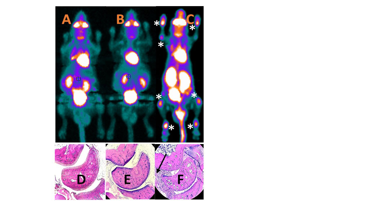
Results: Kv1.3−/− mice did not develop arthritis; whereas. As expected in this model about 75% of the wild-type C57BL/6 mice had arthritis. Arthritic changes appeared around day 28 and lasted beyond day 60. Scores for arthritis (CS and HS) on day-60 was zero in the Kv1.3−/− mice. In WT mice the CS was 2.8 ±0.5 and the HS was 6.2±1.5 (p< .01). In the Figure 1, representative histological sections from ankle joints are shown for Kv1.3−/− and WT mice. Evidence of arthritis was further confirmed/quantified by micro-PET imaging.
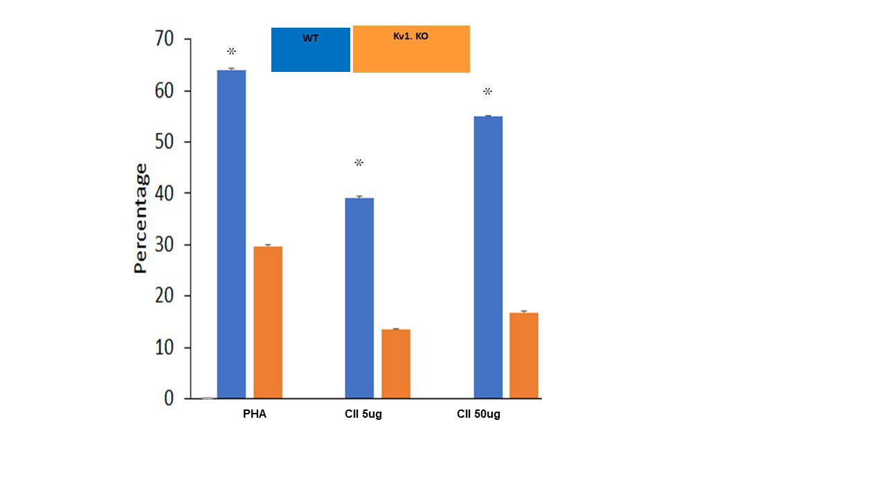
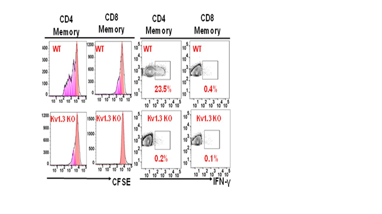
We also observed that knocking out of Kv1.3 renders T cells resistant to activation. As Kv1.3−/− mice did not develop inflammatory arthritis (Fig 1) we hypothesized that the T cells in Kv1.3-/- mice will fail to get activated by CCII. We separated T cells from the spleens of WT and Kv1.3-/- mice on day 60 of CIA induction. Using MTT test and FACS studies we observed a marked proliferation of CD4+ T cells with CCII in the WT compared to the Kv1.3 KO (p< 0.01) (Fig 2); and in the WT mice activation with CCII induced increased CD4+TEM cell proliferative response and secretion of IFN–γ in splenocytes (Fig 3), while in the Kv1.3-/- mice it is clear that knocking out of Kv1.3 renders CD4+ TEM cells resistant to activation (Fig 2 and 3).
Conclusion: In the CIA model here we have demonstrated WT mice developed clinical/histopathological, immunological and radiological evidences of inflammatory arthritis whereas Kv1.3−/− mice did not. This substantiates a critical role of the Kv1.3, a K+ channel in the pathogenesis of T cell mediated autoimmune arthritis including RA. These results provide a platform to develop small molecule-based novel Kv1.3 antagonists (e.g., PAP-1) for treatment of RA.
D, E, F. Histopathological section on day 60 from ankle joints clearly show marked by the black arrow the characteristic histological changes with pannus formation after induction of CIA in the wild mouse (F) whereas in the Kv1.3-KO (E) and in the in the wild type control (D) there was no inflammation or pannus formation following induction of CIA .
To cite this abstract in AMA style:
Raychaudhuri S, Raychaudhuri S, Wulff H. Mice Do Not Develop CIA: Knocking out of Kv1.3 Renders CD4+TEMCells Resistant to Activation [abstract]. Arthritis Rheumatol. 2022; 74 (suppl 9). https://acrabstracts.org/abstract/mice-do-not-develop-cia-knocking-out-of-kv1-3-renders-cd4temcells-resistant-to-activation/. Accessed .« Back to ACR Convergence 2022
ACR Meeting Abstracts - https://acrabstracts.org/abstract/mice-do-not-develop-cia-knocking-out-of-kv1-3-renders-cd4temcells-resistant-to-activation/


