ACR Meeting Abstracts

ACR Meeting Abstracts

  • Meeting Abstracts
    • All Meetings
    • PRSYM 2026
    • ACR Convergence 2025
    • Download Abstract Supplements
  • Keyword Index
  • My Favorites
    • View & print my favorites
  • Search

Home » Meeting Abstracts » ACR Convergence 2025

Abstract Number: 0880

Detecting Interstitial Lung Disease and Identifying Extensive Disease on Chest Computed Tomography in Patients with Systemic Sclerosis: Cut-Offs for Lung Texture Analysis and its Prognostic Implications

Nicholas landini1, lisa Jungblut2, cecilia strappa3, Christian Blüthgen2, Rucsandra Dobrota4, Muriel Elhai5, Carina Mihai6, Sinziana Muraru-Carbune2, Martina Orlandi7, mariaelena Occhipinti8, Khadija El-Aoufy9, Gemma Lepri10, valeria panebianco11, anna rita larici3, Anna-Maria Hoffmann-Vold12, Cosimo Nardi13, Serena Guiducci14, Silvia Bellando-Randone15, Marco Matucci-Cerinic16, Thomas Frauenfelder2, Oliver Distler17 and Cosimo Bruni18, 1Department of Radiological Sciences, Oncology and Pathology, "Sapienza" University, Policlinico Umberto I, Rome, Italy. Department of Rheumatology, University Hospital Zurich, University of Zurich, Zurich, Switzerland, Rueil Malmaison, France, 2Institute for Diagnostic and Interventional Radiology, University Hospital Zurich, University Zurich, Zurich, Switzerland, zurich, Switzerland, 3Advanced Radiology Center, Department of Diagnostic Imaging and Oncological Radiotherapy, Fondazione Policlinico Universitario "A. Gemelli" IRCCS, Rome, Italy, rome, Italy, 4Center of Experimental Rheumatology, Department of Rheumatology, University Hospital Zurich, University of Zurich, the LOOP Zurich, Zurich, Switzerland, 5Institute for Diagnostic and Interventional Radiology, University Hospital Zurich, University Zurich, Zurich, Switzerland, Schlieren, Switzerland, 6University Hospital Zurich, University of Zurich, Zurich, Switzerland, 7Rheumatology Unit, Department of Medical and Surgical Sciences for Children and Adults, University Hospital of Modena and Reggio Emilia School of Medicine, Italy., modena, Italy, 8Division of Radiology, Fondazione Toscana Gabriele Monasterio, Pisa, Italy., pisa, Italy, 9Department of Health Science, University of Florence, Florence, Italy., firenze, Italy, 10Department of Experimental and Clinical Medicine, University of Florence, Division of Rheumatology Scleroderma Unit, AOU Careggi Hospital, Florence, Italy., Florence, Italy, 11Department of Radiological Sciences, Oncology and Pathology, "Sapienza" University, Policlinico Umberto I, Rome, Italy., Rome, Italy, 12Oslo University Hospital, Oslo, Norway, 13Department of Experimental and Clinical Biomedical Sciences, Radiodiagnostic Unit n. 2, University of Florence - Azienda Ospedaliero-Universitaria Careggi, Florence, Italy, Florence, Italy, 14Department of Experimental and Clinical Medicine, Scleroderma Unit, Division of Rheumatology, University of Florence, Azienda Ospedaliera Universitaria Careggi, Florence, Italy, Pistoia, Italy, 15University of Florence, Florence, Florence, Italy, 16University San Raffaele Milano, Milano, Milan, Italy, 17Department of Rheumatology, University Hospital Zurich, University of Zurich, Switzerland, Zurich, Switzerland, 18University of Zurich, Zurich, Switzerland

Share on X (Twitter) Share on Facebook Share on LinkedIn Share on Email
Print

Meeting: ACR Convergence 2025

Keywords: Health Care, Imaging, pulmonary, Scleroderma, Scleroderma, Systemic

Session Information

Date: Monday, October 27, 2025

Title: Abstracts: Systemic Sclerosis & Related Disorders – Clinical II (0879–0884)

Session Type: Abstract Session

Session Time: 10:15AM-10:30AM

Background/Purpose: Interstitial lung disease (ILD) is a leading cause of death in patients with systemic sclerosis (SSc), especially in case of extensive disease detected on computed tomography (CT). Automated post-processing analysis software can compute the extent of ILD through lung texture analysis (LTA) of CT images. However, its use in clinical practice is limited by the lack of standardized thresholds.The aim of our study was to identify and validate optimal thresholds of ILD extent quantified through LTA to detect the presence of ILD and to define extensive ILD in SSc patients.

Methods: SSc patients visiting two SSc referral center between 2005-2021, who underwent chest CT, were included. Technically suitable images (non-contrast, axial inspiratory acquisition, slice thickness and spacing ≤2 mm) were analyzed through LTA (Imbio), quantifying the percentage of lung volume occupied by total ILD (sum of ground glass, reticulation and honeycombing). Two radiologists blinded to the LTA data independently reviewed the CT scans to visually identify the presence of ILD and extensive ILD. The latter was defined as ILD involvement >20% of lung parenchyma or forced vital capacity < 70% predicted in case of indeterminate extent (Goh 2008). Disagreements were resolved by consensus, otherwise through a third reviewer. Patients were randomly split into derivation and validation cohorts using a 2:1 ratio. Receiver operating characteristic (ROC) curves with area under the curve (AUC) were computed to identify the optimal ILD extent threshold for detecting presence of ILD and, in patients with ILD, for identifying extensive disease, using the visual evaluation as the reference standard. Kaplan Meier curves and Cox regression analysis were performed to determine the impact of ILD and extensive disease on both visual and LTA assessments on mortality up to 5 years, the second adjusted for known mortality risk factors (age, sex, diffuse skin subset and pulmonary hypertension).

Results: A total of 664/1118 (58%) SSc patients were eligible for the study (Table 1). Visual analysis identified ILD in 313 (47%) cases, 103 (33%) showing extensive ILD. In the derivation cohort (433 patients, 206 ILD, 38% extensive), ROC analysis identified the optimal total ILD extent threshold for ILD detection at 1% (AUC 0.83) and at 7% for extensive ILD (AUC 0.84). In the validation cohort (231 subjects, 104 ILD, 25% extensive), these thresholds achieved 78% sensitivity / 71% specificity for ILD detection, and 81% sensitivity / 70% specificity for extensive ILD identification. Over median 5 years follow-up, 84 (13%) patients died [37 (12%) in the ILD group]. After adjustment for confounders, both ILD presence (Fig. 1) and extensive disease (Fig. 2) were independent risk factors for mortality, with comparable results by visual and LTA assessments.

Conclusion: Cutoffs for total ILD extent by LTA to detect ILD presence and identify extensive disease were derived and validated, with comparable performance to visual evaluation on negative prognostic impact. Our results lay the foundation for expanding the use of LTA post-processing in SSc-ILD towards automated diagnosis and prognostic stratification.
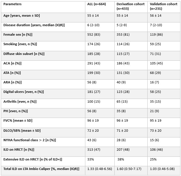
Supporting image 1Table 1. Description of the study population, stratified into derivation and validation cohorts.

ACA: anticentromere antibodies; ARA: anti-RNA polymerase 3 antibodies; ATA: anti-topoisomerase I antibodies; ATA: DLCO: diffusion capacity of the lung for carbon oxide; FVC: forced vital capacity; ILD: interstitial lung disease; IQR: interquartile range; PH: pulmonary hypertension; SD: standard deviation.

Supporting image 2Figure 1. Prediction of mortality for interstitial lung disease identified through visual assessment vs. lung texture analysis.

ILD: interstitial lung disease; HR: hazard ratio; LTA: lung texture analysis.

Supporting image 3Figure 2. Prediction of mortality for extensive (EXT) versus limited (LIM) interstitial lung disease (ILD) identified through visual assessment vs. lung texture analysis (LTA).

EXT: extensive disease; ILD: interstitial lung disease; HR: hazard ratio; LIM: limited disease; LTA: lung texture analysis; PH: pulmonary hypertension.


Disclosures: N. landini: None; l. Jungblut: None; c. strappa: None; C. Blüthgen: Promedical Foundation, 5, Swiss society of radiology, 5; R. Dobrota: Actelion, 5, 6, Amgen, 6, Boehringer-Ingelheim, 6, Iten-Kohaut, 5, Otsuka, 6, Pfizer, 5, Walter und Gertrud Siegenthaler Fellowship, 5; M. Elhai: AstraZeneca, 12, Congress support, Boehringer-Ingelheim, 6, FOREUM, 5, Iten Kohaut foundation, 5, Janssen, 12, Congress support, Kurt und Senta Herrmann foundation, 5, Novartis, 5, Pfizer, 5, University Zurich, 5; C. Mihai: Boehringer Ingelheim, 2, 12, congress support, Janssen, 2, MED Talks Switzerland, 6, Medbase, 6, MedTrix, 6, Mepha, 6, Novartis, 6, PlayToKnow, 6; S. Muraru-Carbune: AstraZeneca, 12, Congress support; M. Orlandi: None; m. Occhipinti: None; K. El-Aoufy: None; G. Lepri: None; v. panebianco: None; a. larici: None; A. Hoffmann-Vold: AbbVie, 2, Avalyn, 2, Boehringer Ingelheim, 2, 5, 6, 12, Medical writing support provided by Fleishman Hillard., Bristol-Myers Squibb, 2, Calluna Pharma, 2, Genentech, 2, Janssen, 2, 5, 6, Medscape, 2, 6, Merck Sharp & Dohme, 2, 6, Novartis, 6, Pliant Therapeutics, 2, Roche, 2, 6, Werfen, 2; C. Nardi: None; S. Guiducci: None; S. Bellando-Randone: Boehringer-Ingelheim, 2, 6; M. Matucci-Cerinic: None; T. Frauenfelder: AstraZeneca, 2, Bayer, 6, Siemens, 6; O. Distler: 4P-Pharma, 2, 6, AbbVie/Abbott, 2, 6, Acceleron, 2, 6, Acepodia Biotech, 2, 6, Aera, 2, 6, AnaMar, 2, 6, Anaveon AG, 2, 6, Argenx, 2, 6, AstraZeneca, 2, 6, BMS, 2, 5, 6, Calluna (Arxx), 2, 6, Cantargia AB, 2, 6, CITUS AG, 8, CSL Behring, 2, 6, EMD Serono, 2, 6, Galapagos, 2, 6, Galderma, 2, 6, Gossamer, 2, 6, Hemetron, 2, 5, 6, Innovaderm, 2, 5, 6, Janssen, 2, 6, Mediar, 2, 5, 6, mir-29 for the treatment of systemic sclerosis, 10, Mitsubishi Tanabe, 2, 5, 6, MSD Merck, 2, 6, Nkarta Inc., 2, 6, Novartis, 2, 6, Orion, 2, 6, Pilan, 2, 6, Prometheus, 2, 6, Quell, 2, 6, Sumitomo, 2, 5, 6, Topadur, 2, 5, 6, UCB, 2, 5, 6; C. Bruni: Boehringer-Ingelheim, 1, 2, 12, Congress support, EMDO foundation, 5, Iten-Kohaut foundation, 5, Novartis foundation for medical-biological research, 5, Scleroderma Clinical Trial Consortium (SCTC), 5, Scleroderma Research Foundation, 5.

To cite this abstract in AMA style:

landini N, Jungblut l, strappa c, Blüthgen C, Dobrota R, Elhai M, Mihai C, Muraru-Carbune S, Orlandi M, Occhipinti m, El-Aoufy K, Lepri G, panebianco v, larici a, Hoffmann-Vold A, Nardi C, Guiducci S, Bellando-Randone S, Matucci-Cerinic M, Frauenfelder T, Distler O, Bruni C. Detecting Interstitial Lung Disease and Identifying Extensive Disease on Chest Computed Tomography in Patients with Systemic Sclerosis: Cut-Offs for Lung Texture Analysis and its Prognostic Implications [abstract]. Arthritis Rheumatol. 2025; 77 (suppl 9). https://acrabstracts.org/abstract/detecting-interstitial-lung-disease-and-identifying-extensive-disease-on-chest-computed-tomography-in-patients-with-systemic-sclerosis-cut-offs-for-lung-texture-analysis-and-its-prognostic-implicatio/. Accessed .

ACR Meeting Abstracts - https://acrabstracts.org/abstract/detecting-interstitial-lung-disease-and-identifying-extensive-disease-on-chest-computed-tomography-in-patients-with-systemic-sclerosis-cut-offs-for-lung-texture-analysis-and-its-prognostic-implicatio/

My Favorites

Save and print abstracts during your browser session by clicking the “Favorite” button at the bottom of any abstract (must have cookies enabled in browser). See saved favorites.

Abstract Policies

  • ACR Convergence Abstract Embargo Policies
  • ACR Convergence Abstract Permissions & Reprints
  • PRSYM Abstract Policies

ACR Convergence. Where Rheumatology Meets

ACR Convergence 2026

Join us November 6-11 in Orlando, Florida.
See Registration Information

  • Contact ACR
  • Privacy Policy
  • ACR Policies
  • Cookie Preferences

© Copyright 2026 American College of Rheumatology