ACR Meeting Abstracts

ACR Meeting Abstracts

  • Meetings
    • ACR Convergence 2024
    • ACR Convergence 2023
    • 2023 ACR/ARP PRSYM
    • ACR Convergence 2022
    • ACR Convergence 2021
    • ACR Convergence 2020
    • 2020 ACR/ARP PRSYM
    • 2019 ACR/ARP Annual Meeting
    • 2018-2009 Meetings
    • Download Abstracts
  • Keyword Index
  • Advanced Search
  • Your Favorites
    • Favorites
    • Login
    • View and print all favorites
    • Clear all your favorites
  • ACR Meetings

Abstract Number: 2531

CRISPR Deletion Screen in Fibroblasts Identifies Novel Regulators of Inflammation

Alisa Mueller1, Suppawat Kongthong2, Angela Zou3, Gerald Watts4, Cassandra Murphy2, Hung Nguyen2, Jacqueline Perrigoue5, Mathew Chamberlain5, Kevin Wei6, Soumya Raychaudhuri2 and Michael Brenner7, 1Brigham and Women's Hospital, Harvard Medical School, Boston, MA, 2Brigham and Women's Hospital, Boston, MA, 3Harvard Medical School, Boston, MA, 4Harvard Medical School, Brookline, MA, 5Johnson & Johnson, Spring House, PA, 6Brigham and Women's Hospital at Harvard Medical School, Boston, MA, 7Brigham and Women's Hospital, Harvard Medical School, Newton, MA

Meeting: ACR Convergence 2024

Keywords: autoimmune diseases, Fibroblasts, Synovial, immunology, Inflammation, rheumatoid arthritis

  • Tweet
  • Click to email a link to a friend (Opens in new window) Email
  • Click to print (Opens in new window) Print
Session Information

Date: Monday, November 18, 2024

Title: Plenary III

Session Type: Plenary Session

Session Time: 9:00AM-10:30AM

Background/Purpose: In rheumatoid arthritis (RA), synovial fibroblasts adopt an inflammatory phenotype that catalyzes pathogenic synovial hyperplasia and ultimately bone and cartilage destruction. However, identifying therapeutic entry points to modulate synovial fibroblast phenotypes remains challenging. Here, we describe the development and execution of an in vitro arrayed CRISPR deletion screen and identify novel molecular regulators of stromal inflammation.

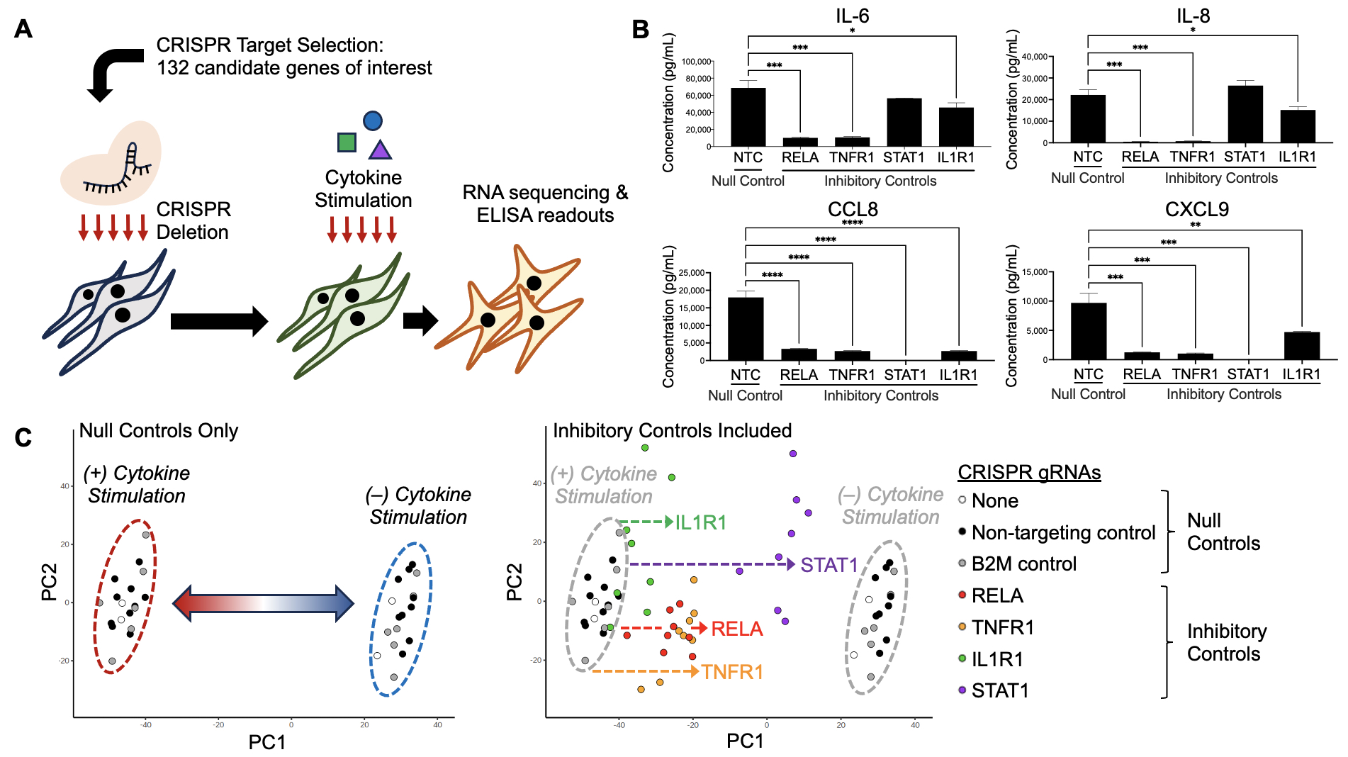
Methods: To generate a custom arrayed CRISPR library for screening, integrated analyses of bulk and single-cell RNA-sequencing (scRNAseq) datasets from OA and RA were used to generate a composite ranking of all genes based on several criteria including expression, upregulation in RA, enrichment in inflammatory fibroblast subsets, fibroblast specificity, and predicted functional relevance. For CRISPR deletion, synovial fibroblasts were nucleofected with guide RNA (gRNA)-Cas9 ribonucleoprotein complexes targeting gene candidates and controls. Cells were cultured for 7 days and stimulated for 16 hours with a combination of TNF-α, IL-17, IFN-γ, and IL-1β, cytokines relevant in RA pathogenesis. Cells and supernatant were processed for downstream bulk RNAseq and ELISA analyses, Western blot, qPCR, and functional assays. Genes upregulated on cytokine stimulation of non-transfected controls were used to develop stromal inflammation signatures.

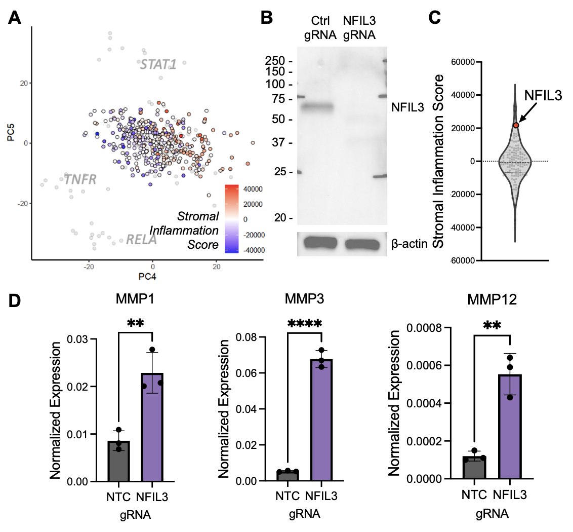
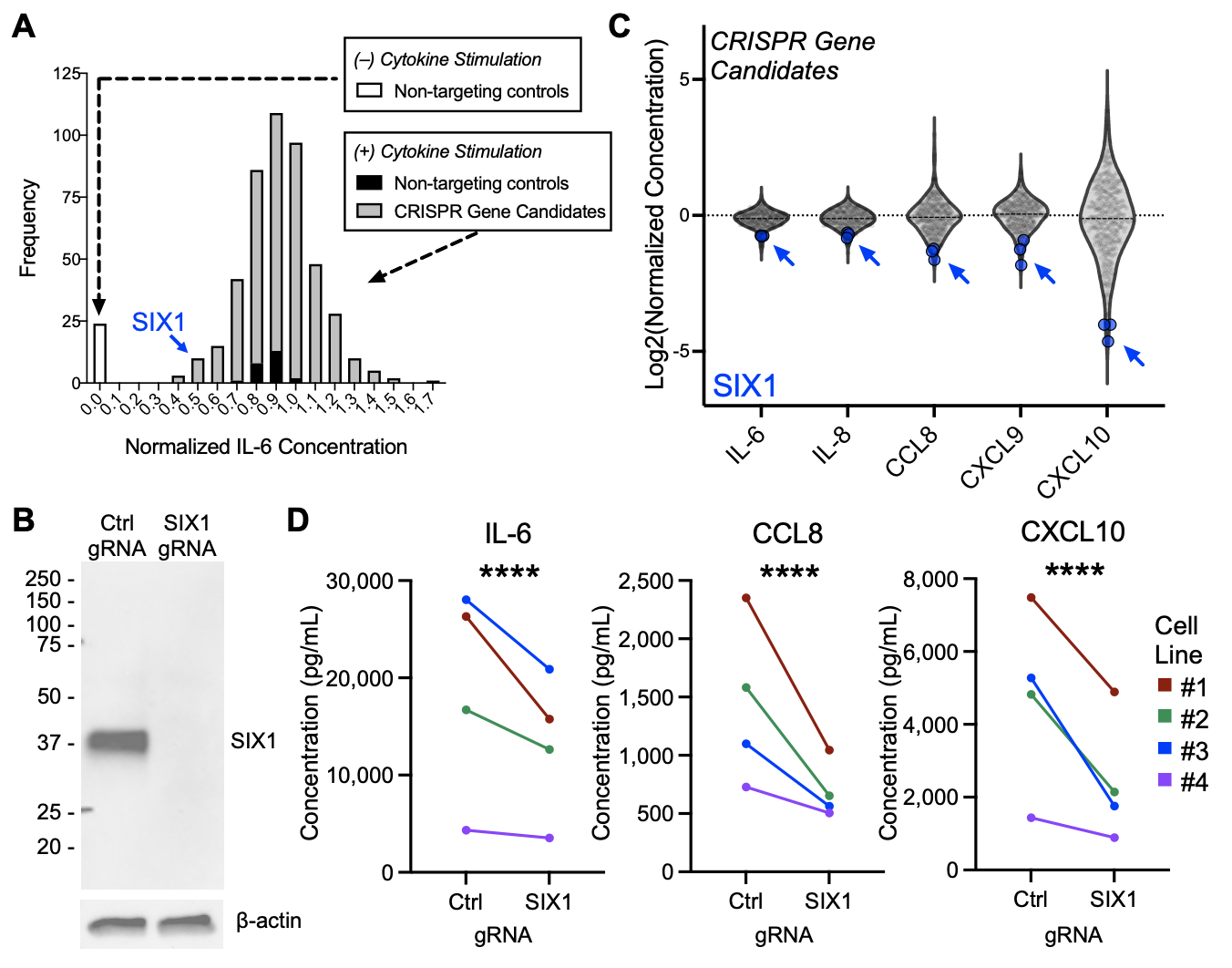
Results: We performed individual CRISPR deletion of 132 genes in an arrayed screen of human RA synovial fibroblasts and evaluated the impact on inflammatory cytokine activation through RNAseq of cell lysates and ELISA of cell culture supernatant (Fig 1A). As a proof-of-concept, we show that deletion of genes known to be active in RA signaling pathways, including RELA, TNFR1, STAT1, and IL1R1, abrogates secretion of pathogenic cytokines (Fig 1B) and attenuates transcriptional changes associated with cytokine stimulation (Fig 1C). Two methods were used for hit calling: one based on enrichment in a stromal inflammation gene signature (Fig 2) and another based on protein expression (Fig 3), and we highlight one hit from each approach. In RNAseq analyses, stromal inflammation signature scores were calculated to identify genes whose CRISPR deletion enhanced or decreased inflammatory potentiation (Fig 2A). Targeting NFIL3 (Fig 2B), a transcription factor implicated in JIA and arthritis severity in mouse models, results in strong enrichment of the inflammation gene signature (Fig 2C) and upregulation of matrix metalloproteases (Fig 2D), suggestive of an invasive phenotype. In protein analyses, the concentrations of ten selected inflammation markers were measured by ELISA to identify candidates that altered cytokine and chemokine secretion (Fig 3A, IL-6 shown). Deletion of SIX1 (Fig 3B), a transcription factor known to promote cell proliferation and tumorigenesis, potently decreased secretion of several cytokines (Fig 3C), which was validated in multiple RA cell lines (Fig 3D).

Conclusion: Here, we show the utility of CRISPR deletion screens to identify novel regulators of fibroblast activation and pathologic effector functions. Hits identified and pathways they regulate may be considered for drug development.

Supporting image 1

Figure 1: Development of a stromal CRISPR deletion screening strategy. A) Schematic of optimized CRISPR screening protocol. For B) and C), the term “null controls” indicates CRISPR conditions that are not expected to have an impact on fibroblast response to cytokine stimulation including the use of no gRNAs, non-targeting gRNAs, and deletion of genes that are predicted not to alter cytokine response (including the gene B2M). The term “inhibitory controls” (labeled by the genes targeted by gRNAs including RELA, TNFR1, STAT1, and IL1R1) indicates CRISPR conditions that are expected to target pathways associated with cytokine stimulation. Hence, their deletion is predicted to inhibit cytokine response. B) Validation of the screening strategy by ELISA measurement of the indicated analytes in synovial fibroblast culture supernatant after stimulation with a combination of cytokines including TNF-α, IL_17, IFN-γ, and IL_1β. Inhibitory controls show decreased cytokine expression in distinct patterns relevant to the control gene targeted for deletion. NTC, non-targeting control. C) Principal component analysis (PCA) plot of RNAseq data. In the left panel, only “null controls” are plotted, and transcriptional changes associated with cytokine activation are shown to separate along the PC1 axis. Non-stimulated conditions are shown with a blue outline and cytokine-stimulated conditions illustrated with a red outline. In the right panel, “inhibitory controls” which have been cytokine-stimulated are overlaid onto the PCA plot. Deletion of inhibitory control genes attenuates transcriptional changes seen with cytokine activation in synovial fibroblasts.

Supporting image 2

Figure 2: Transcriptional analysis reveals novel targetable genes involved in stromal pathogenicity including NFIL3. A) Stromal inflammation scores projected onto a PCA plot of RNA sequencing data from cytokine-activated synovial fibroblasts that have undergone CRISPR deletion of candidates and controls. Each circle represents a single replicate of a candidate gene target or control. A color sliding scale is projected onto candidate genes with red indicating high stromal inflammation signature enrichment and blue indicating decreased enrichment. Controls are labeled in grey with gene names. B) Western blot showing protein knockdown of NFIL3 after gene deletion by CRISPR. C) Violin plot illustrating stromal inflammation scores for each screen target with replicates averaged. The red circle indicates NFIL3, which exhibits one of the highest stromal inflammation scores upon deletion. D) Bar chart showing upregulation of matrix metalloprotease (MMP) transcripts in the setting of NFIL3 gene deletion as detected by qPCR.

Supporting image 3

Figure 3: Cytokine secretion profiles identify genes that alter fibroblast inflammatory potentiation including SIX1. A) Histogram depicting normalized concentrations of IL-6 in supernatant from the CRISPR screen candidates and controls as an example of one of ten analytes measured by ELISA. Replicates are shown for non-targeting controls with no cytokine stimulation (white), non-targeting controls with cytokine stimulation (black), and CRISPR gene candidates (grey). SIX1 CRISPR deletion reduces secreted IL-6 concentration (blue arrow). Log2 transformation of this IL-6 data is depicted in the violin plot in (C) along with other analytes. B) Western blot illustrating knockdown of SIX1 protein with CRISPR deletion. C) Violin plot with ELISA analytes indicated on the x-axis and the concentrations plotted along the y-axis, which are shown as a log2 transformation of the normalized concentrations. Each dot represents an individual replicate of each gene candidate targeted by gRNAs. SIX1 CRISPR deletion is highlighted in blue showing decreased secretion of multiple cytokines. D) Validation of cytokine downregulation with SIX1 CRISPR deletion in synovial fibroblast cell lines from multiple RA patients.


Disclosures: A. Mueller: None; S. Kongthong: None; A. Zou: ; G. Watts: None; C. Murphy: None; H. Nguyen: None; J. Perrigoue: Johnson & Johnson, 3, 11; M. Chamberlain: Gilead, 11, Janssen, 3, 11, Pfizer, 11; K. Wei: 10X Genomics, 5, Gilead sciences, 2, Merck/MSD, 5, Mestag, 2, santa ana bio, 2; S. Raychaudhuri: Janssen, 1, Mestag, 8, Nimbus, 2, Pfizer, 1, Sonoma, 8, Third Rock Ventures, 2; M. Brenner: GlaxoSmithKlein(GSK), 2, Mestag Therapeutics, 2, 11, Moderna, 2.

To cite this abstract in AMA style:

Mueller A, Kongthong S, Zou A, Watts G, Murphy C, Nguyen H, Perrigoue J, Chamberlain M, Wei K, Raychaudhuri S, Brenner M. CRISPR Deletion Screen in Fibroblasts Identifies Novel Regulators of Inflammation [abstract]. Arthritis Rheumatol. 2024; 76 (suppl 9). https://acrabstracts.org/abstract/crispr-deletion-screen-in-fibroblasts-identifies-novel-regulators-of-inflammation/. Accessed .
  • Tweet
  • Click to email a link to a friend (Opens in new window) Email
  • Click to print (Opens in new window) Print

« Back to ACR Convergence 2024

ACR Meeting Abstracts - https://acrabstracts.org/abstract/crispr-deletion-screen-in-fibroblasts-identifies-novel-regulators-of-inflammation/

Advanced Search

Your Favorites

You can save and print a list of your favorite abstracts during your browser session by clicking the “Favorite” button at the bottom of any abstract. View your favorites »

All abstracts accepted to ACR Convergence are under media embargo once the ACR has notified presenters of their abstract’s acceptance. They may be presented at other meetings or published as manuscripts after this time but should not be discussed in non-scholarly venues or outlets. The following embargo policies are strictly enforced by the ACR.

Accepted abstracts are made available to the public online in advance of the meeting and are published in a special online supplement of our scientific journal, Arthritis & Rheumatology. Information contained in those abstracts may not be released until the abstracts appear online. In an exception to the media embargo, academic institutions, private organizations, and companies with products whose value may be influenced by information contained in an abstract may issue a press release to coincide with the availability of an ACR abstract on the ACR website. However, the ACR continues to require that information that goes beyond that contained in the abstract (e.g., discussion of the abstract done as part of editorial news coverage) is under media embargo until 10:00 AM ET on November 14, 2024. Journalists with access to embargoed information cannot release articles or editorial news coverage before this time. Editorial news coverage is considered original articles/videos developed by employed journalists to report facts, commentary, and subject matter expert quotes in a narrative form using a variety of sources (e.g., research, announcements, press releases, events, etc.).

Violation of this policy may result in the abstract being withdrawn from the meeting and other measures deemed appropriate. Authors are responsible for notifying colleagues, institutions, communications firms, and all other stakeholders related to the development or promotion of the abstract about this policy. If you have questions about the ACR abstract embargo policy, please contact ACR abstracts staff at [email protected].

Wiley

  • Online Journal
  • Privacy Policy
  • Permissions Policies
  • Cookie Preferences

© Copyright 2025 American College of Rheumatology