Session Information
Date: Monday, October 27, 2025
Title: (0934–0954) Systemic Lupus Erythematosus – Animal Models Poster
Session Type: Poster Session B
Session Time: 10:30AM-12:30PM
Background/Purpose: Autoimmune disease is far more common in women than men. We have determined that the skin of healthy women is primed for autoimmunity due to increased baseline epidermal expression of the transcription coregulator VGLL3. Accordingly, VGLL3 overexpression in transgenic (TG) mouse epidermis causes cutaneous and systemic murine lupus. Intriguingly, VGLL3 is also reported to act variably as an oncogene and tumor suppressor, revealing pleiotropic roles. While fibrosis is a hallmark of multiple autoimmune skin diseases, few animal models successfully recapitulate fibrosis, limiting mechanistic investigation.
Methods: Trichrome staining, immunostaining, qPCR, bulk RNA-seq, and single-cell RNA-seq (scRNA-seq) were used to analyze skin from mouse models of epidermal VGLL3 overexpression and human skin from patients with cutaneous lupus and healthy controls. Seurat, IPA, CellChat, and CellPhoneDB were used for analysis of scRNA-seq data. Human keratinocytes overexpressing VGLL3 and primary human skin fibroblasts were used for conditioned media experiments.
Results: We demonstrate that epidermal VGLL3 also drives autoimmune skin fibrosis. Integrating single-cell transcriptomic data from TG mice and human lupus patients, we establish a role for VGLL3-overexpressing keratinocytes in initiating autoimmune skin fibrosis through altering fibroblast transcriptional states. In pre-lesional skin, VGLL3-overexpressing keratinocytes upregulate specific cytokines including TNF and CCL20 and promote an interferon-rich environment, inducing pre-lesional fibroblasts to activate a profibrotic, proinflammatory transcriptional program implicated in fibrotic disease. Conditioned media experiments confirm a direct effect of keratinocytes on fibroblasts, increasing transcription of CCL2, a chemokine implicated in multiple fibrotic diseases. Analysis of fibroblasts reveals expansion in pre-lesional and lesional skin of a population of profibrotic fibroblasts that are shown by ligand-receptor analysis to be one of the most differentially activated cellular communicators in diseased skin. Fibroblast subclustering identifies a transgenic mouse-specific cluster of Tnc-positive fibroblasts, which have been shown to be crucial in the progression and maintenance of fibrotic skin diseases by promoting fibroblast activation and sustaining fibrotic responses. Analysis of human cutaneous lupus scRNA-seq data corroborates many of our murine lupus findings in human disease.
Conclusion: These results suggest that VGLL3 and keratinocytes play a dual role in early autoimmune skin fibrosis through both fibroblast reprogramming and immune cell recruitment and support the use of the K5-Vgll3 model for investigation of autoimmune skin fibrosis.
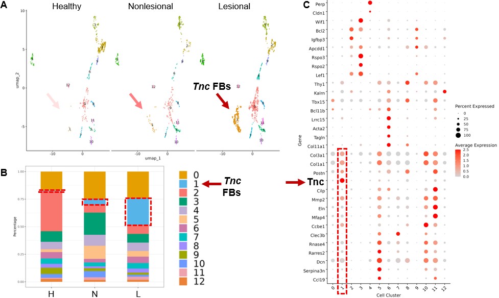 Figure 1. Epidermal VGLL3 overexpression leads to autoimmune skin fibrosis.
A. Gross images of female and male transgenic (TG) and wild-type (WT) mice with epidermal overexpression of Vgll3.
B. H&E staining of murine lesional TG and WT skin and human lupus patient and healthy control skin.
C. Trichrome staining of murine and human skin as in (B).
D. CTGF/CCN2 immunohistochemistry (IHC) of murine and human skin as in (B).
.jpg) Figure 2. Single-cell RNA-sequencing (scRNA-seq) reveals immune cell expansion in skin lesions induced by epidermal VGLL3 overexpression
Figure 2. Single-cell RNA-sequencing (scRNA-seq) reveals immune cell expansion in skin lesions induced by epidermal VGLL3 overexpression
A. Uniform Manifold Approximation and Projections (UMAPs) of all cells colored by cell type in healthy (H), nonlesional transgenic (N), and lesional transgenic (L) mouse skin.
B. Dot plot of representative marker genes for each cell type.
C. Histogram of all cells colored by cell type across disease states. Red boxes, immune cells.
.jpg) Figure 3. Fibroblast subclustering reveals emergence of Tnc-expressing fibroblasts in nonlesional and lesional transgenic mouse skin.
Figure 3. Fibroblast subclustering reveals emergence of Tnc-expressing fibroblasts in nonlesional and lesional transgenic mouse skin.
A. Fibroblast subclustering across disease states. Red arrows indicate Tnc fibroblasts (FBs) (cells in orange).
B. Histogram of fibroblast subclusters across disease states. Red boxes indicate Tnc fibroblasts (bars in light blue).
C. Dot plot of representative marker genes for each cell type. Red boxes indicate Tnc fibroblasts.
To cite this abstract in AMA style:
Gharaee-Kermani M, Dey P, van Drongelen V, Rew J, Bogle R, Hildebrandt M, Klein B, Moallemian R, Verhaegen M, Dlugosz A, Tsoi L, Kahlenberg J, Gudjonsson J, Billi A. The Cancer-associated Female-biased Factor VGLL3 Drives Autoimmunity and Fibrosis [abstract]. Arthritis Rheumatol. 2025; 77 (suppl 9). https://acrabstracts.org/abstract/the-cancer-associated-female-biased-factor-vgll3-drives-autoimmunity-and-fibrosis/. Accessed .« Back to ACR Convergence 2025
ACR Meeting Abstracts - https://acrabstracts.org/abstract/the-cancer-associated-female-biased-factor-vgll3-drives-autoimmunity-and-fibrosis/