ACR Meeting Abstracts

ACR Meeting Abstracts

  • Meetings
    • ACR Convergence 2024
    • ACR Convergence 2023
    • 2023 ACR/ARP PRSYM
    • ACR Convergence 2022
    • ACR Convergence 2021
    • ACR Convergence 2020
    • 2020 ACR/ARP PRSYM
    • 2019 ACR/ARP Annual Meeting
    • 2018-2009 Meetings
    • Download Abstracts
  • Keyword Index
  • Advanced Search
  • Your Favorites
    • Favorites
    • Login
    • View and print all favorites
    • Clear all your favorites
  • ACR Meetings

Abstract Number: 1755

Unique Pattern of Cadherin 6 Localization in Fibroblast-Like Synoviocytes

Camilla R. L. Machado1, David Boyle1, Narayanan B. Perumal2, Robert J. Benschop3 and Gary Firestein1, 1University of California San Diego, San Diego, CA, 2Eli Lilly and Company, San Diego, CA, 3Eli Lilly and Company, Indianapolis, IN

Meeting: ACR Convergence 2023

Keywords: autoimmune diseases, Epigenetics, Fibroblasts, Synovial, Inflammation, rheumatoid arthritis

  • Tweet
  • Click to email a link to a friend (Opens in new window) Email
  • Click to print (Opens in new window) Print
Session Information

Date: Tuesday, November 14, 2023

Title: (1734–1775) RA – Etiology and Pathogenesis Poster

Session Type: Poster Session C

Session Time: 9:00AM-11:00AM

Background/Purpose: The synovial lining of rheumatoid arthritis (RA) is formed by a network of fibroblast-like synoviocytes (FLS). Cadherins are type I transmembrane proteins and participate in FLS cell-to-cell contacts, particularly cadherin 11. Unbiased profiling of cadherin expression in FLS using epigenetic data identified cadherin 6 (CDH6) as a gene of interest in RA FLS. CDH6, which is a type II cadherin, has mainly been studied in oncology where high expression is associated with cancer progression and invasion. CDH6 in FLS could regulate similar functions in RA. To understand the diverse functions of CDH6 in FLS, we analyzed its cellular distribution in synovial tissue.

Methods: RA and osteoarthritis (OA) synovial tissue (n=6 each) were obtained at arthroplasty. RA FLS and OA FLS lines (n=6 each) were derived from enzymatically dispersed synovial tissue and used from passages 5-7. Western blot analysis was used for protein quantification with anti-CDH6 antibody and was normalized to GAPDH. The distribution of CDH6 in FLS and synovial tissue was evaluated by immunofluorescence and confocal microscopy.

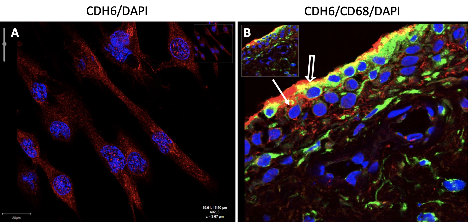
Results: Initial immunofluorescence studies of RA synovial tissue revealed abundant CDH6 in the intimal lining. Using anti-CD68 and anti-CDH6 antibodies in double stained tissues, we observed CDH6 expression in lining macrophages and fibroblasts. CDH6 was also expressed to a lesser degree in the sublining, particularly in the perivascular regions and included macrophages and non-macrophages (Figure 1). A similar distribution of CDH6 protein was observed in OA synovium and suggests that CDH6 is an important component in the synovial architecture. Because FLS reside in the intimal lining and were strongly CDH6 positive, we explored the expression and distribution of CDH6 in cultured FLS. Western blots showed that expression is higher in RA than OA FLS (1.2±0.01 vs 0.1±0.01 OD, p=0.03; n=4 each). Using immunostaining, expression was higher in FLS cultured at higher cell density and was ~2-fold higher in confluent vs. non-confluent cultures. Thus, CDH6 might be regulated, in part, by cell-cell contact. More intriguing, confocal microscopy showed significant expression of CDH6 protein in the cytoplasm, peri-nuclear regions, and nucleus in addition to the cell membrane.

Conclusion: CDH6 is expressed in synovial lining and sublining fibroblasts and macrophages. The striking intracellular distribution suggests additional functions beyond adhesion and homotypic aggregation, such as signalling and regulation of gene transcription. Thus, CDH6 has similarities to E-Cadherin, which also localizes to intracellular structures and participates in biologic functions. Our data suggest for the first time that CDH6 might have multiple functions in FLS that contribute to its pathogenic behaviour.

Supporting image 1

Figure 1. CDH6 expression in RA FLS and RA synovium. We performed confocal microscopy to determine if CDH6 is present in the synovial lining and its distribution in RA FLS. A) Representative image of RA FLS with anti-CDH6 (red) and DAPI (blue). CDH6 is found in the cytoplasm, peri-nuclear regions, nucleus, and the cell membrane. B) Representative image of RA synovium with anti-CDH6 (red) and -CD68 (green) (macrophage) antibodies show lining and sublining expression of CDH6, most likely in FLS (solid white arrow) and lining macrophages (open white arrow).


Disclosures: C. R. L. Machado: Eli Lilly, 5; D. Boyle: None; N. B. Perumal: Eli Lilly, 3; R. J. Benschop: Eli Lilly and Company, 3, 11; G. Firestein: Eli Lilly, 5.

To cite this abstract in AMA style:

R. L. Machado C, Boyle D, B. Perumal N, J. Benschop R, Firestein G. Unique Pattern of Cadherin 6 Localization in Fibroblast-Like Synoviocytes [abstract]. Arthritis Rheumatol. 2023; 75 (suppl 9). https://acrabstracts.org/abstract/unique-pattern-of-cadherin-6-localization-in-fibroblast-like-synoviocytes/. Accessed .
  • Tweet
  • Click to email a link to a friend (Opens in new window) Email
  • Click to print (Opens in new window) Print

« Back to ACR Convergence 2023

ACR Meeting Abstracts - https://acrabstracts.org/abstract/unique-pattern-of-cadherin-6-localization-in-fibroblast-like-synoviocytes/

Advanced Search

Your Favorites

You can save and print a list of your favorite abstracts during your browser session by clicking the “Favorite” button at the bottom of any abstract. View your favorites »

All abstracts accepted to ACR Convergence are under media embargo once the ACR has notified presenters of their abstract’s acceptance. They may be presented at other meetings or published as manuscripts after this time but should not be discussed in non-scholarly venues or outlets. The following embargo policies are strictly enforced by the ACR.

Accepted abstracts are made available to the public online in advance of the meeting and are published in a special online supplement of our scientific journal, Arthritis & Rheumatology. Information contained in those abstracts may not be released until the abstracts appear online. In an exception to the media embargo, academic institutions, private organizations, and companies with products whose value may be influenced by information contained in an abstract may issue a press release to coincide with the availability of an ACR abstract on the ACR website. However, the ACR continues to require that information that goes beyond that contained in the abstract (e.g., discussion of the abstract done as part of editorial news coverage) is under media embargo until 10:00 AM ET on November 14, 2024. Journalists with access to embargoed information cannot release articles or editorial news coverage before this time. Editorial news coverage is considered original articles/videos developed by employed journalists to report facts, commentary, and subject matter expert quotes in a narrative form using a variety of sources (e.g., research, announcements, press releases, events, etc.).

Violation of this policy may result in the abstract being withdrawn from the meeting and other measures deemed appropriate. Authors are responsible for notifying colleagues, institutions, communications firms, and all other stakeholders related to the development or promotion of the abstract about this policy. If you have questions about the ACR abstract embargo policy, please contact ACR abstracts staff at [email protected].

Wiley

  • Online Journal
  • Privacy Policy
  • Permissions Policies
  • Cookie Preferences

© Copyright 2025 American College of Rheumatology