ACR Meeting Abstracts

ACR Meeting Abstracts

  • Meetings
    • ACR Convergence 2024
    • ACR Convergence 2023
    • 2023 ACR/ARP PRSYM
    • ACR Convergence 2022
    • ACR Convergence 2021
    • ACR Convergence 2020
    • 2020 ACR/ARP PRSYM
    • 2019 ACR/ARP Annual Meeting
    • 2018-2009 Meetings
    • Download Abstracts
  • Keyword Index
  • Advanced Search
  • Your Favorites
    • Favorites
    • Login
    • View and print all favorites
    • Clear all your favorites
  • ACR Meetings

Abstract Number: 2599

Xist Deletion in B Cells Results in Systemic Lupus Erythematosus Phenotypes

Claudia Lovell1, Nikhil Jiwrajka2, Hayley Amerman3, Michael Cancro4 and Montserrat Anguera5, 1Perelman School of Medicine at the University of Pennsylvania, Philadelphia, PA, 2Hospital of the University of Pennsylvania, Philadelphia, PA, 3University of Pennsylvania School of Veterinary Medicine, Philadelphia, PA, 4Perelman School of Medicine, Philadelphia, PA, 5University of Pennsylvania, Philadelphia, PA

Meeting: ACR Convergence 2024

Keywords: Autoantibody(ies), B-Lymphocyte, Epigenetics, lupus-like disease, Non-coding RNA

  • Tweet
  • Click to email a link to a friend (Opens in new window) Email
  • Click to print (Opens in new window) Print
Session Information

Date: Monday, November 18, 2024

Title: Abstracts: SLE – Etiology & Pathogenesis

Session Type: Abstract Session

Session Time: 3:00PM-4:30PM

Background/Purpose: Systemic lupus erythematosus (SLE) exhibits a strong female sex bias but the mechanisms underlying this bias are not well understood. Epidemiological studies demonstrate that SLE risk increases with the number of X chromosomes carried by an individual, suggesting important genetic contributions from the X chromosome. To ensure dosage compensation of X-linked genes relative to XY males, mammals with multiple X chromosomes utilize X-chromosome inactivation (XCI), a multi-layered epigenetic mechanism that is initiated and maintained by the long non-coding RNA Xist, which is expressed from and coats the inactive X chromosome. The X chromosome is enriched for genes with immunomodulatory functions that are relevant to B cell function. B cells from female SLE patients and mouse models of SLE exhibit mislocalization of Xist RNA and aberrant expression of X-linked genes, suggesting that impairment of XCI may contribute to disease. We sought to determine whether perturbed XCI maintenance in the B cell compartment contributes to SLE pathogenesis.

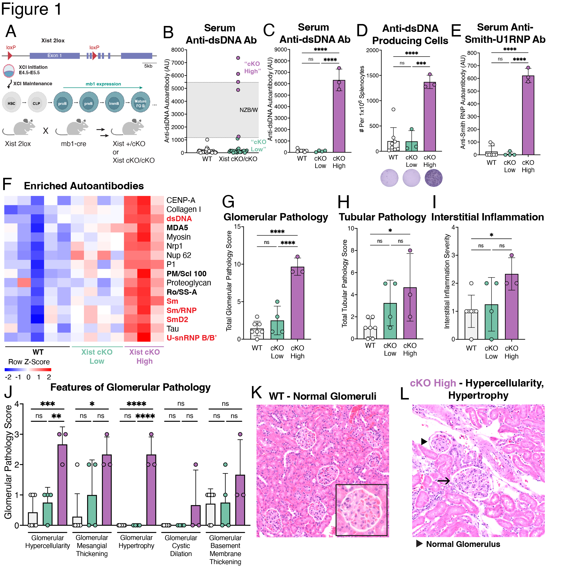
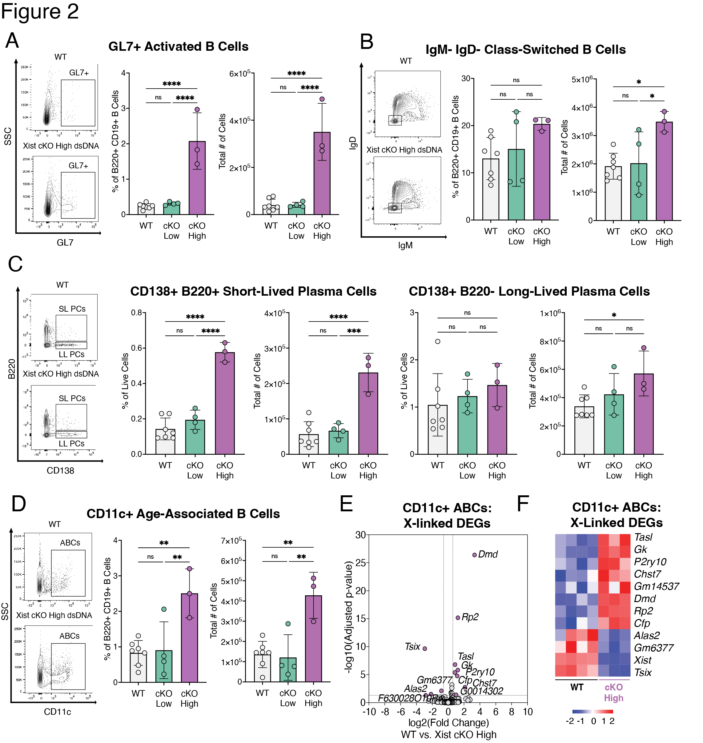
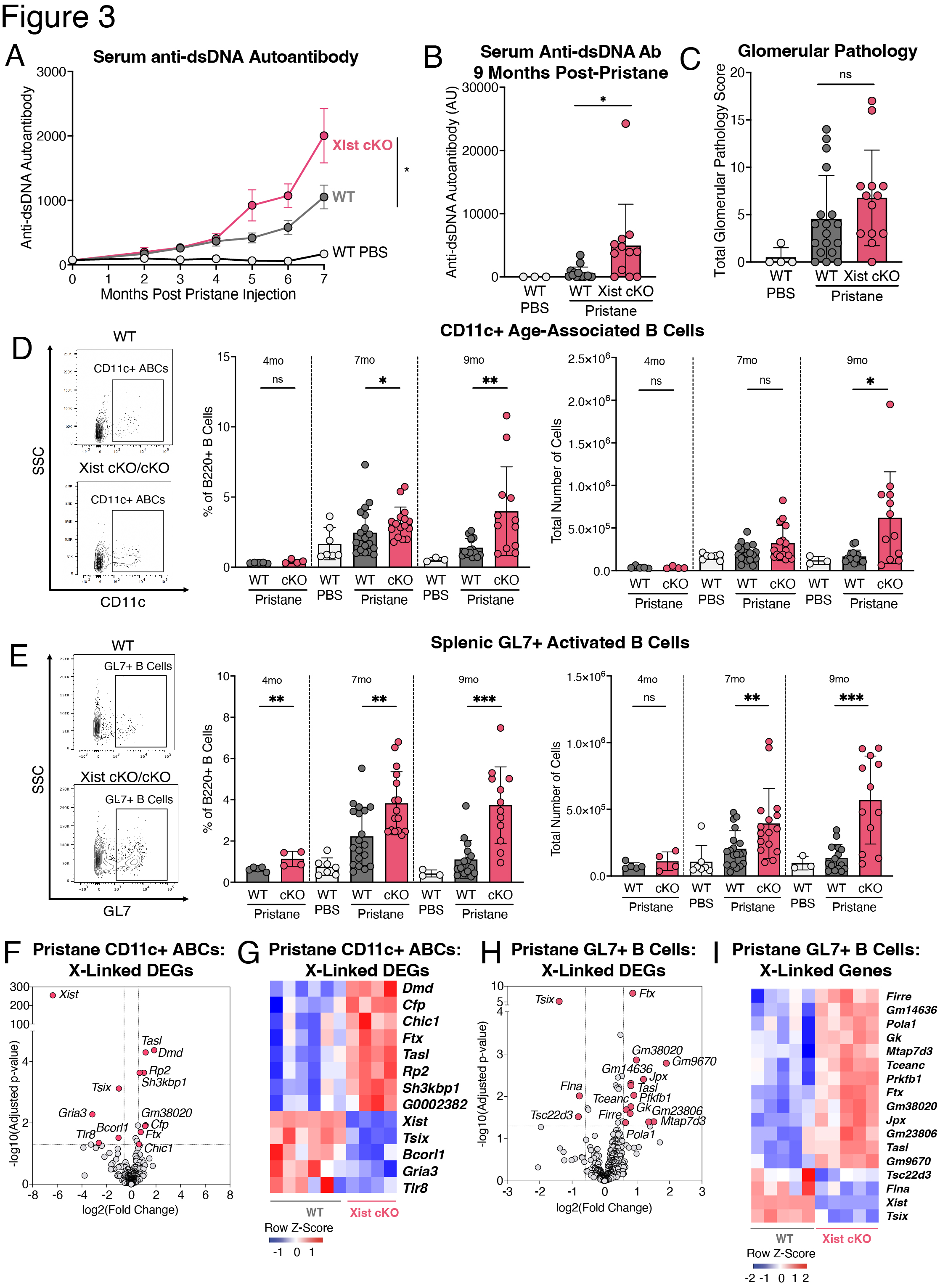
Methods: We generated female BALB/c mice with a B cell specific deletion of Xist (mb1WT/creXistfl/fl, “Xist cKO”). We measured serum levels of anti-dsDNA autoantibodies in a cohort of WT (n=109) and Xist cKO (n=66) mice. In a cohort of Xist cKO mice with high anti-dsDNA (n=3 “Xist cKO High”), Xist cKO mice with low anti-dsDNA (n=4), and WT controls (n=7) we measured sera for various autoantibodies, quantified splenic B cell subsets, and assessed kidney pathology. We also injected a cohort of WT (n=46) and Xist cKO mice (n=36) with pristane to induce SLE-like disease and measured anti-dsDNA autoantibodies, kidney pathology, and splenic B cells during disease. We performed RNAseq on activated B cells from Xist cKO mice with spontaneous and pristane-induced SLE.

Results: Some Xist cKO mice spontaneously developed elevated anti-dsDNA, anti-Smith U1-RNP, anti-MDA5, anti-PM/Scl-100, and anti-Ro/SS-A, as well as glomerular kidney pathology. These Xist cKO mice also had more activated B cell subsets including GL7+ activated B cells, class-switched B cells, age-associated B cells (ABCs), and plasma cells. Pristane-treated Xist cKO mice developed higher levels of anti-dsDNA, had slightly more severe glomerular kidney pathology, and had more activated B cell subsets compared to pristane-treated WT mice. Activated B cells from Xist cKO mice with spontaneous and pristane-induced SLE demonstrated upregulation of the X-linked TLR7 pathway member Tasl.

Conclusion: Our data demonstrate that perturbed XCI maintenance via Xist deletion in the B cell compartment results in the upregulation of X-linked Tasl, a contributor to abnormal immune signaling in SLE, along with activated B cell subset expansion, production of disease-specific autoantibodies, and glomerulonephritis. These findings suggest that disruption of XCI maintenance in B cells can both spontaneously confer and exacerbate SLE-like disease, thereby providing a novel pathogenic mechanism that simultaneously accounts for the strong female sex bias of SLE.

Supporting image 1

A. Breeding schematic used to generate mice with a B cell-specific Xist deletion. Xist 2lox mice with loxP sites flanking the first 3 exons of Xist were successively bred to mb1-cre mice for deletion in the B cell lineage staring with early proB cells, to generate mice with a homozygous Xist deletion (“Xist cKO/cKO”) or a heterozygous Xist deletion (“Xist +/cKO”) in the B cell compartment. B. Serum levels of anti-dsDNA autoantibodies from female BALB/c mice 12 months or older (n=39 female WT mice, n=27 female Xist cKO/cKO mice) measured by enzyme-linked immunosorbent assay (ELISA). Each circle represents an individual mouse. Grey bar bounded by dotted lines represents serum levels of anti-dsDNA in NZB/W (n=8) mice that spontaneously develop SLE-like disease. C. Mean serum anti-dsDNA autoantibody levels ± standard deviation (SD) in female Xist cKO/cKO mice with elevated anti-dsDNA autoantibodies (“Xist cKO High”, n=3), age-matched female Xist cKO/cKO mice with low anti-dsDNA autoantibodies (“Xist cKO Low”, n=4), and age-matched female WT controls (“WT”, n=7) as measured by ELISA. D. Mean number of anti-dsDNA producing cells per 1,000,000 splenocytes ± SD as measured by ELISPOT. E. Mean serum anti-Smith RNP autoantibody levels ± SD as measured by ELISA. F. Heatmap depicting individual sample-level data for those autoantibodies significantly enriched in Xist cKO High mice compared to WT mice or Xist cKO Low mice, as measured by an autoantigen microarray. Bold autoantibodies in black are either specific for or associated with non-SLE female biased autoimmune diseases. G. Mean total glomerular pathology ± SD. H. Mean total tubular pathology ± SD. I. Mean interstitial inflammation ± SD. J. Mean scores ± SD for individual features of glomerular pathology. K. Representative image of glomeruli from a WT mouse. L. Representative image of glomeruli from Xist cKO High mice. * p < 0.05, ** p < 0.01, *** p < 0.001, **** p < 0.0001 via ordinary one-way ANOVA with Tukey’s multiple comparisons test.

Supporting image 2

A-D. Cells were pre-gated on live single cells that are CD4-CD8-Gr_1-F4/80-. Quantification of splenic (A) GL7+ activated B cells (B220+CD19+IgM-IgD-CD138-GL7+), (B) class-switched B cells (B220+CD19+IgM-IgD-), (C) short-lived plasma cells (SLPCs; IgD-CD138+B220+) and long-lived plasma cells (LLPCs; IgD-CD138+B220-), and (D) age-associated B cells (ABCs; B220+CD19+CD11c+). Data are depicted as mean ± SD. * p < 0.05, ** p < 0.01, *** p < 0.001, **** p < 0.0001 via ordinary one-way ANOVA with Tukey’s multiple comparisons test. E. Volcano plot depicting X-linked genes that are significantly differentially expressed (purple) between Xist cKO High and WT CD11c+ ABCs. F. Heatmap of differentially expressed X-linked genes between Xist cKO High and WT CD11c+ ABCs.

Supporting image 3

A. Mean serum anti-dsDNA autoantibody levels ± SEM in pristane-treated Xist cKO mice (n=32), pristane-treated WT mice (n=41), and PBS-treated WT mice (n=10) over 7 months. p < 0.05 by mixed effects model with the Geisser-Greenhouse correction (not assuming equal variability of differences). B. Mean serum anti-dsDNA autoantibody levels ± SD in pristane-treated Xist cKO mice (n=12), pristane-treated WT mice (n=18), and PBS-treated WT mice (n=3) at 9 months post-pristane injection. C. Mean total glomerular pathology score + SD. D-E. Cells were pre-gated on live, single CD4-CD8-Gr_1-F4/80- cells. Quantification of splenic (D) age-associated B cells (ABCs; B220+CD19+CD11c+) and (E) GL7+ activated B cells (B220+CD19+IgM-IgD-CD138-GL7+) after 4, 7, or 9 months of pristane. Data are depicted as mean + SD. * p < 0.05, ** p < 0.01, *** p < 0.001 via pairwise Mann-Whitney U test comparing pristane-treated Xist cKO to pristane-treated WT mice. F. Volcano plot depicting X-linked genes that are significantly differentially expressed (pink) in CD11c+ ABCs from pristane-treated Xist cKO vs. pristane-treated WT mice. G. Heatmap of differentially expressed X-linked genes in CD11c+ ABCs from pristane-treated mice. H. Volcano plot depicting X-linked genes that are significantly differentially expressed (pink) in GL7+ activated B cells from pristane-treated Xist cKO vs. pristane-treated WT mice. I. Heatmap of differentially expressed X-linked genes in GL7+ B cells from pristane-treated mice.


Disclosures: C. Lovell: None; N. Jiwrajka: None; H. Amerman: None; M. Cancro: None; M. Anguera: None.

To cite this abstract in AMA style:

Lovell C, Jiwrajka N, Amerman H, Cancro M, Anguera M. Xist Deletion in B Cells Results in Systemic Lupus Erythematosus Phenotypes [abstract]. Arthritis Rheumatol. 2024; 76 (suppl 9). https://acrabstracts.org/abstract/xist-deletion-in-b-cells-results-in-systemic-lupus-erythematosus-phenotypes/. Accessed .
  • Tweet
  • Click to email a link to a friend (Opens in new window) Email
  • Click to print (Opens in new window) Print

« Back to ACR Convergence 2024

ACR Meeting Abstracts - https://acrabstracts.org/abstract/xist-deletion-in-b-cells-results-in-systemic-lupus-erythematosus-phenotypes/

Advanced Search

Your Favorites

You can save and print a list of your favorite abstracts during your browser session by clicking the “Favorite” button at the bottom of any abstract. View your favorites »

All abstracts accepted to ACR Convergence are under media embargo once the ACR has notified presenters of their abstract’s acceptance. They may be presented at other meetings or published as manuscripts after this time but should not be discussed in non-scholarly venues or outlets. The following embargo policies are strictly enforced by the ACR.

Accepted abstracts are made available to the public online in advance of the meeting and are published in a special online supplement of our scientific journal, Arthritis & Rheumatology. Information contained in those abstracts may not be released until the abstracts appear online. In an exception to the media embargo, academic institutions, private organizations, and companies with products whose value may be influenced by information contained in an abstract may issue a press release to coincide with the availability of an ACR abstract on the ACR website. However, the ACR continues to require that information that goes beyond that contained in the abstract (e.g., discussion of the abstract done as part of editorial news coverage) is under media embargo until 10:00 AM ET on November 14, 2024. Journalists with access to embargoed information cannot release articles or editorial news coverage before this time. Editorial news coverage is considered original articles/videos developed by employed journalists to report facts, commentary, and subject matter expert quotes in a narrative form using a variety of sources (e.g., research, announcements, press releases, events, etc.).

Violation of this policy may result in the abstract being withdrawn from the meeting and other measures deemed appropriate. Authors are responsible for notifying colleagues, institutions, communications firms, and all other stakeholders related to the development or promotion of the abstract about this policy. If you have questions about the ACR abstract embargo policy, please contact ACR abstracts staff at [email protected].

Wiley

  • Online Journal
  • Privacy Policy
  • Permissions Policies
  • Cookie Preferences

© Copyright 2025 American College of Rheumatology