
Session Information
Date: Monday, November 18, 2024
Title: T Cell Biology & Targets in Autoimmune & Inflammatory Disease Poster
Session Type: Poster Session C
Session Time: 10:30AM-12:30PM
Background/Purpose: Approximately 80% of cutaneous lupus erythematosus (CLE) patients experience sensitivity to ultraviolet (UV) sunlight rays, which leads to disfiguring skin lesions or systemic disease flares. The mechanisms underlying photosensitivity in CLE remain unknown, but it is well established that both at baseline and in response to UV CLE patients have a high skin type I interferon (IFN) signature. Though mucosal-associated invariant T (MAIT) cells can be activated in a TCR-independent fashion by cytokines, including IFNs, their role in the skin response to UV is unknown.
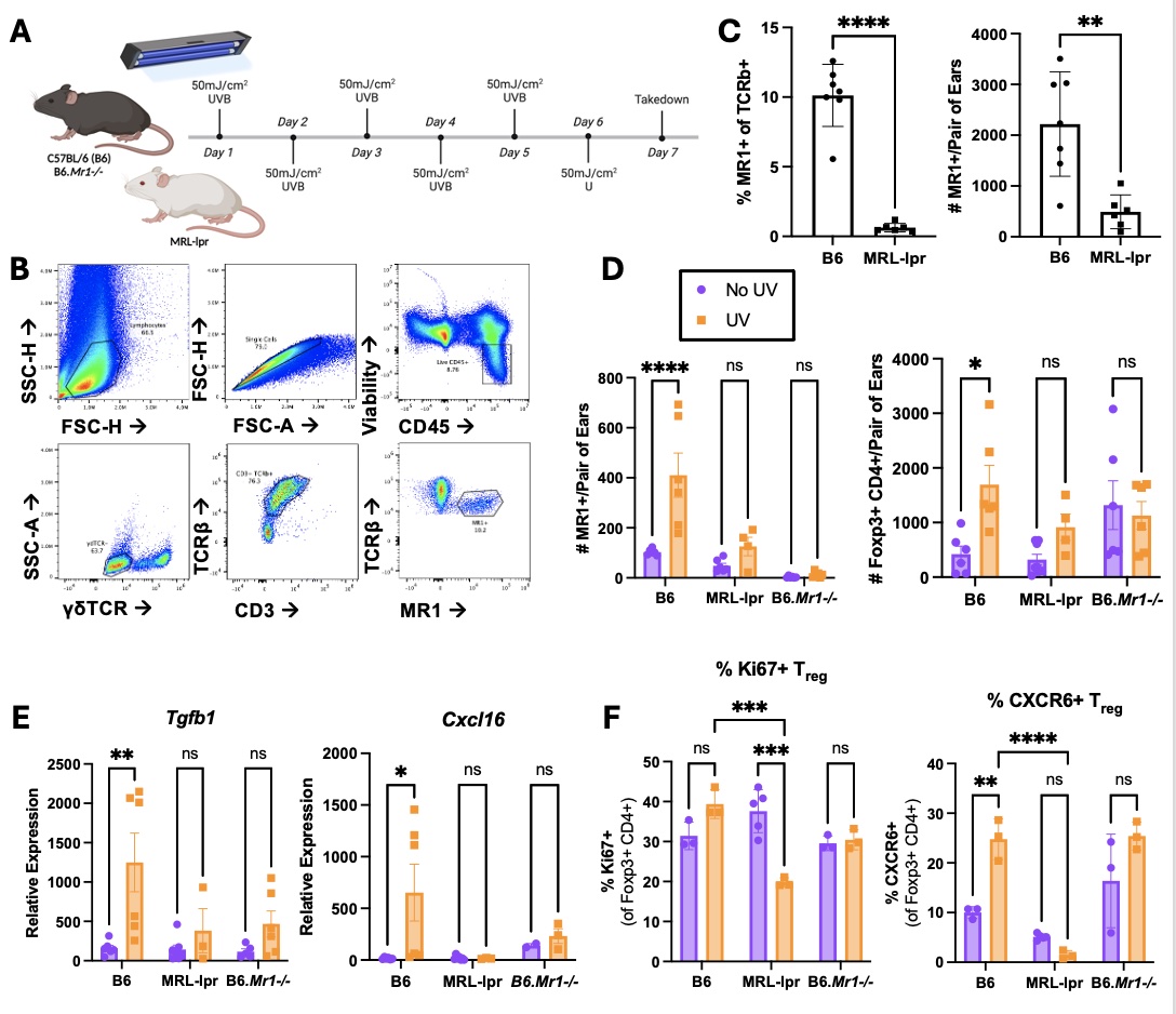
Methods: 8-week old female control (B6 and Mrl/MpJ), lupus (Mrl/MpJ-lpr, Faslpr< ![if !supportAnnotations] >[SG1]< ![endif] > ), MAIT-deficient (B6.Mr1-/-), and IFNAR-deficient (B6. Ifnar-/-) mice were exposed to a chronic course of low-dose UVB (50mJ/cm2 daily, 6 total doses; Fig. 1A). Differences in skin inflammatory responses and immune cells, including MAIT detection by 5-OP-RU-loaded MR1 tetramer staining (Fig. 1B) and Treg proliferation and phenotype, were quantified by qPCR and flow cytometry, respectively.
Results: At baseline, young pre-lesional MRL-lpr mice harbor ~10-fold less MAIT cells in the skin compared to age-matched B6 mice (Fig. 1B-C). In healthy (B6) skin, exposure to UVB resulted in expansion of MAIT cells with concurrent expansion of regulatory T cells (Treg) compared to unexposed controls (Fig. 1D). However, in MRL-lpr skin, MAIT cells and Treg both failed to expand following UV. UV-induced Treg expansion was also absent in MAIT-deficient (B6.Mr1-/-) skin. In B6 skin, UV exposure stimulated Tgfb1 and Cxcl16 expression, but strikingly this induction was absent in MRL-lpr and B6.Mr1-/- skin (Fig. 1E). Furthermore, in B6 skin after UV, more Treg expressed tissue retention Cxcl16 receptor CXCR6 and proliferation marker Ki67, which was not observed in MRL-lpr or B6.Mr1-/- skin (Fig. 1F). Findings in age-matched Mrl/MpJ skin closely mirrored those in B6 skin. Using IFNAR-deficient mice, we showed that IFN-I signaling is required for UV-induced MAIT expansion in B6 skin.
Conclusion: In healthy skin, concurrent MAIT cell and Treg expansion after UV exposure supports a protective role for MAIT cells in skin responses to UV. UV-induced Treg proliferation and retention are MAIT-cell dependent and may be mediated via Tgfb1 and Cxcl16 induction after UV. These mechanisms are absent in Mrl-lpr skin and may be a consequence of low MAIT cell levels at baseline. We are currently investigating why MAIT cells are almost absent from Mrl-lpr skin and whether MAIT expansion can prevent UV-induced skin inflammation and injury in this lupus model.
To cite this abstract in AMA style:
Crossland G, Mendyka L, Dowling K, Constantinides M, Skopelja-Gardner S. Uncovering a MAIT-Treg Axis in Skin UV Response: Implications for Photosensitive Reactions in Cutaneous Lupus Erythematosus [abstract]. Arthritis Rheumatol. 2024; 76 (suppl 9). https://acrabstracts.org/abstract/uncovering-a-mait-treg-axis-in-skin-uv-response-implications-for-photosensitive-reactions-in-cutaneous-lupus-erythematosus/. Accessed .« Back to ACR Convergence 2024
ACR Meeting Abstracts - https://acrabstracts.org/abstract/uncovering-a-mait-treg-axis-in-skin-uv-response-implications-for-photosensitive-reactions-in-cutaneous-lupus-erythematosus/