ACR Meeting Abstracts

ACR Meeting Abstracts

  • Meetings
    • ACR Convergence 2024
    • ACR Convergence 2023
    • 2023 ACR/ARP PRSYM
    • ACR Convergence 2022
    • ACR Convergence 2021
    • ACR Convergence 2020
    • 2020 ACR/ARP PRSYM
    • 2019 ACR/ARP Annual Meeting
    • 2018-2009 Meetings
    • Download Abstracts
  • Keyword Index
  • Advanced Search
  • Your Favorites
    • Favorites
    • Login
    • View and print all favorites
    • Clear all your favorites
  • ACR Meetings

Abstract Number: 1845

Treg Expansion and IL-6 Induced STAT3 Phosphorylation in CD4+ T Cells Is a Biomarker of Disease Flare in Rheumatoid Arthritis

Amy Anderson1, Luke Jones2, Fiona Rayner3, Jessica Swift1, Daniel Maunder1, Henrique de Paula Lemos1, David Swan4, Abbie Degnan1, Imogen Wilson1, Julie Diboll1, Gary Reynolds1, Jasmine Sim1, Andrew Melville5, Stefan Siebert5, Iain McInnes6, Carl Goodyear5, Catharien Hilkens1, Karim Raza7, Christopher Buckley8, Kenneth Baker1, Arthur Pratt3, Andrew Filer7 and John Isaacs1, 1Translational and Clinical Research Institute, NIHR Newcastle Biomedical Research Centre, Newcastle University and The Newcastle Upon Tyne Hospitals NHS Foundation Trust, Newcastle upon Tyne, United Kingdom, 2Rheumatology Research Group, Institute for Inflammation and Ageing, NIHR Birmingham Biomedical Research Center and Clinical Research Facility, Birmingham, United Kingdom, 3Translational and Clinical Research Institute, NIHR Newcastle Biomedical Research Centre, Newcastle University and The Newcastle Upon Tyne Hospitals NHS Foundation Trust, Newcastle upon Tyne, England, United Kingdom, 4Translational and Clinical Research Institute, NIHR Newcastle Biomedical Research Centre, Newcastle University and The Newcastle Upon Tyne Hospitals NHS Foundation Trust, Newcastle upon Tyne, 5School of Infection and Immunity, College of Medical, Veterinary and Life Sciences, University of Glasgow, Glasgow, United Kingdom, 6University of Glasgow, College of Medical Veterinary and Life Sciences, Glasgow, United Kingdom, 7Rheumatology Research Group, Institute for Inflammation and Ageing, NIHR Birmingham Biomedical Research Center and Clinical Research Facility, University of Birmingham, Birmingham, United Kingdom, 8Kennedy Institute of Rheumatology, University of Oxford, Oxford, United Kingdom

Meeting: ACR Convergence 2024

Keywords: cytokines, rheumatoid arthritis, T Cell, Treg cells

  • Tweet
  • Click to email a link to a friend (Opens in new window) Email
  • Click to print (Opens in new window) Print
Session Information

Date: Monday, November 18, 2024

Title: T Cell Biology & Targets in Autoimmune & Inflammatory Disease Poster

Session Type: Poster Session C

Session Time: 10:30AM-12:30PM

Background/Purpose: Understanding the mechanisms that drive disease flares in patients with rheumatoid arthritis (RA) may aid in the development of biomarkers to facilitate targeted treatment withdrawal in patients in remission and may suggest therapeutic targets for flare prevention. However, these mechanisms are difficult to study due to the unpredictable nature of flare. The BIOlogical Factors that Limit sustAined Remission in rhEumatoid arthritis (BIO-FLARE) study1 is an experimental medicine study of ‘synchronised’ RA flares in which treatment is withdrawn in RA patients in clinical remission receiving conventional synthetic (cs)DMARDs. Subsequently, approximately 50% flare within 6 months, the remainder maintaining drug-free remission (DFR). We have assessed, longitudinally, circulating T cell subsets and mediators following treatment cessation, to better under the mechanisms driving flare and maintaining DFR.

Methods: Individuals with RA receiving csDMARDs, in clinician-defined remission with a DAS28-CRP < 2.4, were recruited into BIO-FLARE. Treatment was stopped and participants monitored over 24 weeks, with serum and Smart Tube-fixed peripheral blood stored at 0, 2, 5, 8, 12 and 24 weeks. Flare was defined as DAS28-CRP ≥3.2 at any study visit, or DAS28-CRP ≥2.4 on two occasions within a 14-day period. Circulating T cell subsets were assessed using a 16 marker Phosflow panel. Soluble serum mediators were detected by the NULISAseq™ Inflammation Panel 2502.

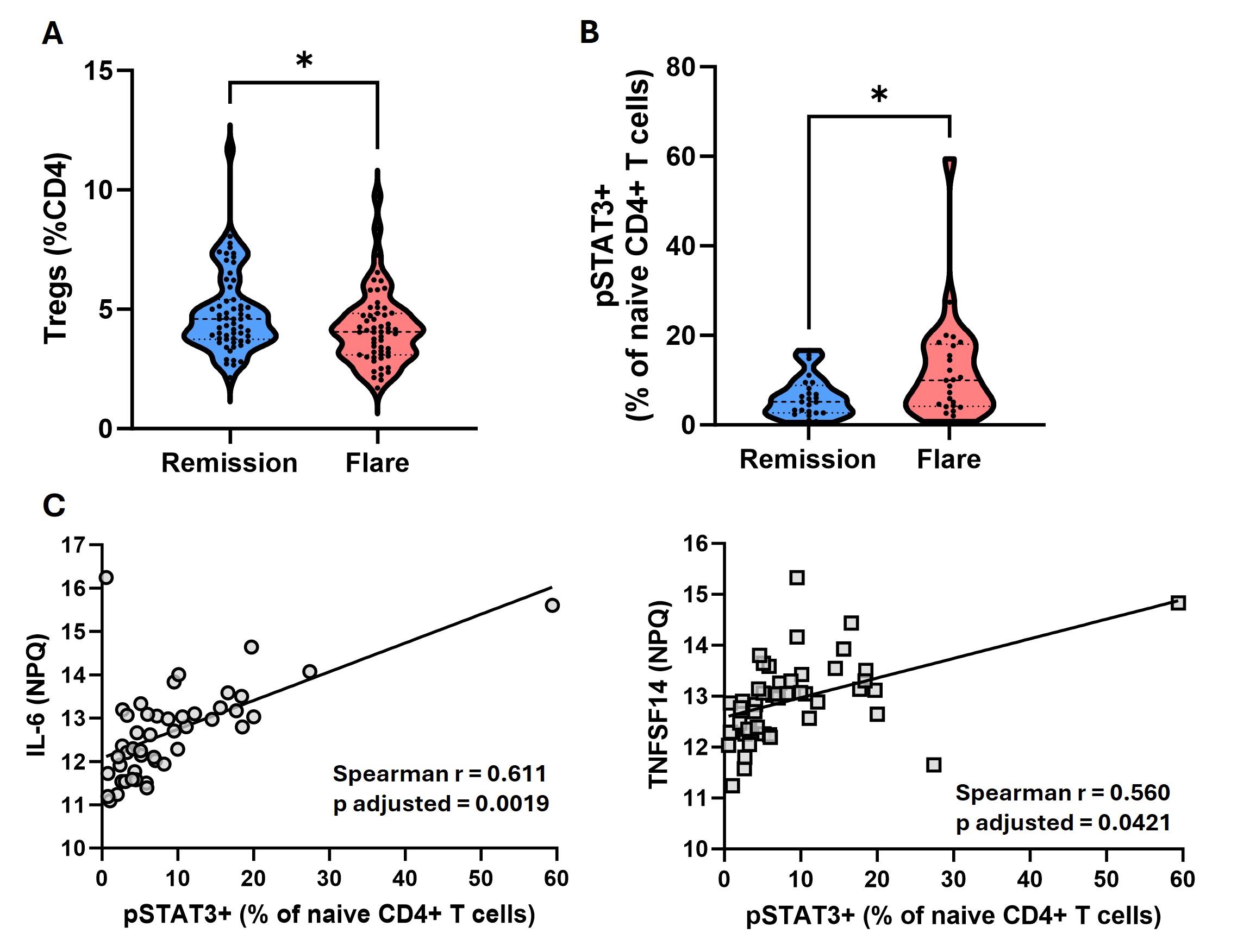
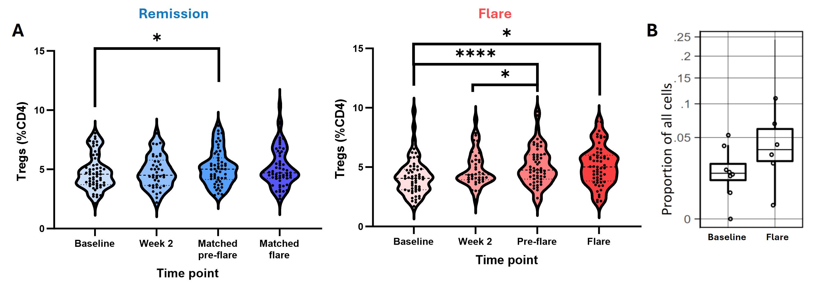
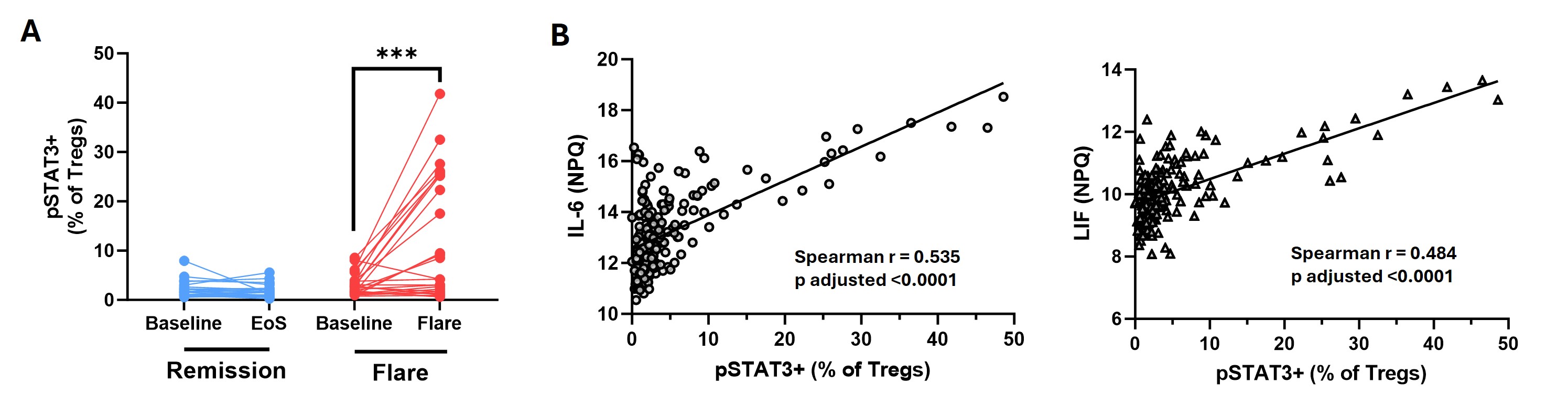
Results: At baseline, participants who subsequently flared and those who maintained DFR were well matched clinically, except for RF/ACPA titres, which were higher in those who subsequently flared. In contrast, flow cytometry analysis at baseline revealed an increase in the proportion of CD4+CD25high Tregs in the DFR group, as well as an increase in the proportion of naive CD4+ T cells expressing phosphorylated STAT3 (pSTAT3) in the flare group (Figure 1A-B). The proportion of pSTAT3+ naive CD4+ T cells strongly correlated with baseline levels of IL-6 and TNFSF14 (Figure 1C), despite similar levels of these cytokines in the two groups at baseline. When assessing longitudinal changes following DMARD withdrawal, Tregs increased in both groups (Figure 2A), as well as in the synovium of flare patients (Figure 2B). Expression of pSTAT3 in Tregs increased over time in the flare group only (Figure 3A) and correlated with IL-6 and LIF levels (Figure 3B).

Conclusion: Even at baseline there are elevated proportions of naive CD4+ T cells expressing pSTAT3 in patients who subsequently flare, and increased Treg proportions in those who achieve DFR. Whilst Treg proportions increase after DMARD withdrawal in both groups, those who flare have increased pSTAT3 expression in this subset, possibly driven by elevated circulating IL-6 and LIF. As pSTAT3 expression is suggested to impair Treg function, the Tregs expanded during flare may be dysfunctional due to their exposure to IL-6 and other pro-inflammatory cytokines. This work highlights key pathways involved in disease flare that warrant further investigation.

References

  1. Rayner F et al. BMC Rheumatol. 2021
  2. Feng W et al. Nat Commun. 2023
  3. Yang L et al. J Dermatol Sci. 2016

Supporting image 1

Figure 1: Tregs are increased at baseline in DFR, whereas STAT3 activation in naïve CD4+ T cells is increased in flare at baseline and correlates with IL-6 and TNFSF14 levels. T cell subsets (Tregs (CD4+CD25high cells) (A) and pSTAT3+ naïve CD4 T cells (B)) were measured in Smart Tube stored peripheral blood from baseline using Phosflow. * p< 0.05 using a Mann Whitney U test. Serum proteins were detected using the NULISAseq™ Inflammation Panel 250. C. Correlation of IL-6 (left) and TNFSF14 (right) with pSTAT3+ naïve CD4 T cells at baseline was assessed using a Spearman correlation with Bonferroni correction. In A remission n = 59 and flare n = 57. In B-C remission n = 25 and flare n = 25.

Supporting image 2

Figure 2: Treg proportions increase over time from the point of DMARD withdrawal in both remission and flare and increase in the synovium of flare patients at the point of flare. A. Tregs (CD4+CD25high T cells) were measured in Smart Tube stored peripheral blood from remission patients (left) and flare patients (right) at 4 different time points following DMARD cessation using Phosflow. B. Tregs in the synovium were assessed using single-cell RNA-Seq at baseline and the point of flare. * p< 0.05, **** p < 0.0001 using a Friedman test with a Dunn’s post-test. In A remission n = 59 and flare n = 57. In B n = 9.

Supporting image 3

Figure 3: pSTAT3 expression in Tregs increases towards the point of flare following DMARD withdrawal and correlates with levels of serum IL-6 and LIF. Tregs (CD4+CD25high T cells) expressing pSTAT3 were measured in Smart Tube stored peripheral blood using Phosflow (A). Serum proteins were detected using the NULISAseq™ Inflammation Panel 250. B. Correlation of IL-6 (left) and LIF (right) with pSTAT3+ Tregs at all time points were assessed using a Spearman correlation with Bonferroni correction. * p< 0.05 using a Wilcoxon signed-rank test. n = 25 and flare n = 25.


Disclosures: A. Anderson: None; L. Jones: None; F. Rayner: None; J. Swift: None; D. Maunder: None; H. de Paula Lemos: None; D. Swan: None; A. Degnan: None; I. Wilson: None; J. Diboll: None; G. Reynolds: None; J. Sim: None; A. Melville: None; S. Siebert: AbbVie, 6, Amgen, 6, AstraZeneca, 6, Boehringer-Ingelheim, 5, Bristol-Myers Squibb, 5, Eli Lilly, 5, GlaxoSmithKline, 5, Janssen, 5, 6, Teijin Pharma, 6, UCB, 5; I. McInnes: AbbVie, 2, 5, 6, Amgen, 2, Bristol Myers Squibb, 2, 5, Cabaletta, 2, Celgene, 2, Compugen, 2, Dextera, 2, Eli Lilly, 2, 5, Janssen, 2, 5, Moonlake, 2, Novartis, 2, 5, Pfizer, 2, UCB, 2, 5, 6; C. Goodyear: None; C. Hilkens: None; K. Raza: None; C. Buckley: None; K. Baker: Pfizer, 5; A. Pratt: None; A. Filer: None; J. Isaacs: AbbVie/Abbott, 2, 6, AnaptysBio, 2, Annexon, 2, AstraZeneca, 2, Bristol-Myers Squibb(BMS), 2, Dragonfly, 2, Eli Lilly, 1, Galapagos, 2, GlaxoSmithKlein(GSK), 2, 5, Istesso, 2, Janssen, 5, Kira Biotech, 2, Ono Pharma, 2, Pfizer, 5, Revelo, 2, Sanofi, 2, Sonoma Biotherapeutics, 2, Topas, 2, UCB, 1.

To cite this abstract in AMA style:

Anderson A, Jones L, Rayner F, Swift J, Maunder D, de Paula Lemos H, Swan D, Degnan A, Wilson I, Diboll J, Reynolds G, Sim J, Melville A, Siebert S, McInnes I, Goodyear C, Hilkens C, Raza K, Buckley C, Baker K, Pratt A, Filer A, Isaacs J. Treg Expansion and IL-6 Induced STAT3 Phosphorylation in CD4+ T Cells Is a Biomarker of Disease Flare in Rheumatoid Arthritis [abstract]. Arthritis Rheumatol. 2024; 76 (suppl 9). https://acrabstracts.org/abstract/treg-expansion-and-il-6-induced-stat3-phosphorylation-in-cd4-t-cells-is-a-biomarker-of-disease-flare-in-rheumatoid-arthritis/. Accessed .
  • Tweet
  • Click to email a link to a friend (Opens in new window) Email
  • Click to print (Opens in new window) Print

« Back to ACR Convergence 2024

ACR Meeting Abstracts - https://acrabstracts.org/abstract/treg-expansion-and-il-6-induced-stat3-phosphorylation-in-cd4-t-cells-is-a-biomarker-of-disease-flare-in-rheumatoid-arthritis/

Advanced Search

Your Favorites

You can save and print a list of your favorite abstracts during your browser session by clicking the “Favorite” button at the bottom of any abstract. View your favorites »

All abstracts accepted to ACR Convergence are under media embargo once the ACR has notified presenters of their abstract’s acceptance. They may be presented at other meetings or published as manuscripts after this time but should not be discussed in non-scholarly venues or outlets. The following embargo policies are strictly enforced by the ACR.

Accepted abstracts are made available to the public online in advance of the meeting and are published in a special online supplement of our scientific journal, Arthritis & Rheumatology. Information contained in those abstracts may not be released until the abstracts appear online. In an exception to the media embargo, academic institutions, private organizations, and companies with products whose value may be influenced by information contained in an abstract may issue a press release to coincide with the availability of an ACR abstract on the ACR website. However, the ACR continues to require that information that goes beyond that contained in the abstract (e.g., discussion of the abstract done as part of editorial news coverage) is under media embargo until 10:00 AM ET on November 14, 2024. Journalists with access to embargoed information cannot release articles or editorial news coverage before this time. Editorial news coverage is considered original articles/videos developed by employed journalists to report facts, commentary, and subject matter expert quotes in a narrative form using a variety of sources (e.g., research, announcements, press releases, events, etc.).

Violation of this policy may result in the abstract being withdrawn from the meeting and other measures deemed appropriate. Authors are responsible for notifying colleagues, institutions, communications firms, and all other stakeholders related to the development or promotion of the abstract about this policy. If you have questions about the ACR abstract embargo policy, please contact ACR abstracts staff at [email protected].

Wiley

  • Online Journal
  • Privacy Policy
  • Permissions Policies
  • Cookie Preferences

© Copyright 2025 American College of Rheumatology