ACR Meeting Abstracts

ACR Meeting Abstracts

  • Meetings
    • ACR Convergence 2024
    • ACR Convergence 2023
    • 2023 ACR/ARP PRSYM
    • ACR Convergence 2022
    • ACR Convergence 2021
    • ACR Convergence 2020
    • 2020 ACR/ARP PRSYM
    • 2019 ACR/ARP Annual Meeting
    • 2018-2009 Meetings
    • Download Abstracts
  • Keyword Index
  • Advanced Search
  • Your Favorites
    • Favorites
    • Login
    • View and print all favorites
    • Clear all your favorites
  • ACR Meetings

Abstract Number: 2539

Telocytes Integrated into Mast Cells and Joint-Draining Lymphatic Vessels Potentially Regulate Lymphatic Clearance

Yue Peng1, H. Mark Kenney2, Karen Bentley1, Lianping Xing3, Benjamin Korman4, Christopher Ritchlin5 and Edward Schwarz1, 1University of Rochester School of Medicine and Dentistry, Rochester, NY, 2University of Rochester School of Medicine and Dentistry, Henrietta, NY, 3University of Rochester School of Medicine and Dentistry, Webster, NY, 4University of Rochester, Rochester, NY, 5Department of Medicine, Allergy, Immunology, and Rheumatology Division, University of Rochester Medical School, Canandaigua, NY

Meeting: ACR Convergence 2024

Keywords: Animal Model, Fibroblasts, Other, Fibroblasts, Synovial, rheumatoid arthritis, Tumor necrosis factor (TNF)

  • Tweet
  • Click to email a link to a friend (Opens in new window) Email
  • Click to print (Opens in new window) Print
Session Information

Date: Monday, November 18, 2024

Title: Abstracts: RA – Animal Models

Session Type: Abstract Session

Session Time: 1:00PM-2:30PM

Background/Purpose: Rheumatoid arthritis (RA) patients and TNF-transgenic mice have lymphatic dysfunction (1). Recently, we showed mast cells involvement, as genetic ablation and drug inhibition decreased lymphatic drainage and exacerbated arthritis in TNF-Tg mice (2). Single-cell RNA sequencing studies identified genes (i.e., Efhd1) selectively expressed in popliteal lymphatic vessels (PLVs) (3). However, the location and function of Efhd1–expressing cells and their relationship with mast cells are not known. Here we aimed to characterize a novel Efhd1-CreERT2 mouse model as a tool for PLV gain and loss-of-function studies and elucidate the mechanisms of peri-lymphatic mast cell regulation.


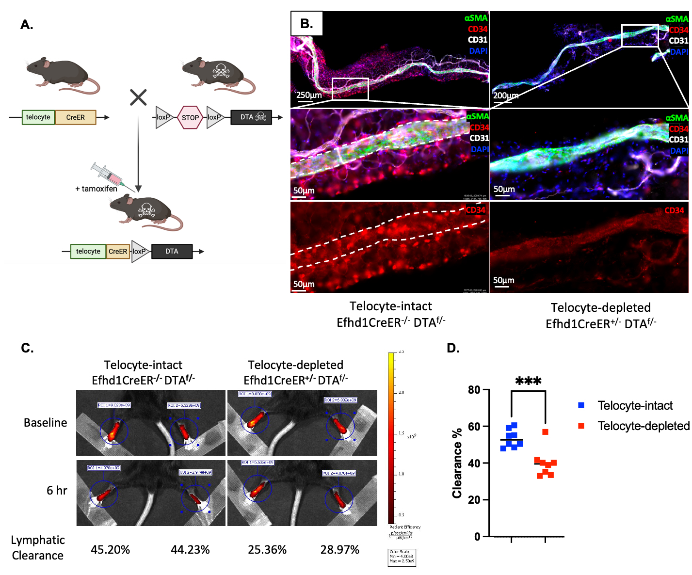
Methods: Efhd1-CreERT2 mice were crossed to Ai9-tdTomato (tdT) reporter mice. The heterozygous offspring were treated with tamoxifen as neonates or adults for lineage tracing studies in the popliteal vasculature and adjacent adipose tissue via immunofluorescent microscopy (IFM) with subsequent transmission electron microscopy (TEM).  The Efhd1-CreERT2 mice were also crossed to ROSA-diphtheria toxin A (DTA) mice to determine in vivo cell depletion efficiency and lymphatic clearance by IFM and near-infrared imaging of indocyanine green (NIR-ICG) injected into the footpad, respectively.


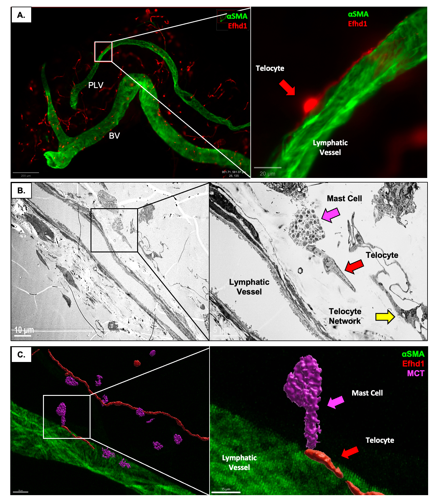
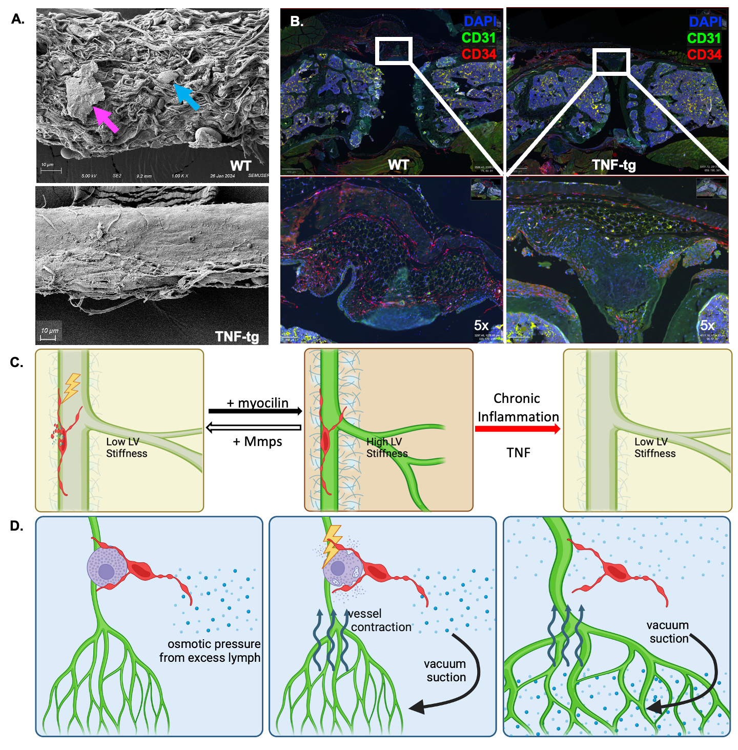
Results: Developmental and adult lineage tracing demonstrated Efhd1-tdT+ labeling of peri-vascular telocytes based on their fibroblastic morphology and characteristic telopods (Figure 1A). Telocyte identity was confirmed by CD31–/CD34+/Vimentin+ IFM (4), and histomorphometry revealed their proportion in WT synovium (84.15%±7.99%) was significantly decreased in TNF-Tg synovium (26.04%±7.38%; p< 0.001) (Figure 3B). Parallel TEM (Figure 1B) and IFM (Figure 1C) revealed tdT+ telopods integrated along the PLV, and within mast cells indicating direct cytoplasmic connections between these cells. DTA-induced depletion of telocytes resulted in reduced lymphatic clearance (Figure 2), suggesting that telocytes may mediate mast cell and PLV communication to regulate lymphatic function.


Conclusion: We developed and validated a novel Efhd1-CreERT2 transgenic mouse for inducible gain and loss-of-function studies in telocytes, whose function in joint homeostasis and arthritis is unknown.  Two distinct subtypes of peri-PLV telocytes exist: one with telopods longitudinally attached along PLVs, and the other with telopods integrated within the plasma membrane of mast cells. Based on these findings, we hypothesize that: 1) PLV telocytes sense and modulate the extracellular matrix (ECM) stiffness of PLVs, potentially through the secretion of myocilin, TIMPs and MMPs (3) for ECM remodeling (Figure 3A&C); and 2) interstitial telocytes that may sense osmotic pressure and regulate PLV contractility, potentially by interacting with mast cells to induce the release of factors (e.g. histamine) that modulate PLV contractions (Figure 3D). Our findings are also consistent with the loss of telocytes in RA synovium (4), phenotypic changes to a yet to be defined population of fibroblastic cells in RA synovium (5) that may be telocytes, and lymphatic dysfunction during disease progression (1).

Supporting image 1

Figure 1. Novel Efhd1-reporter mice reveal direct telocyte-mast cell interactions and telocytes embedded along lymphatic vessels. (A) Immunofluorescent image of a popliteal lymphatic vessel (PLV) from a tamoxifen-treated Efhd1-CreERT2 x Ai9-tdTomato (tdT) mouse stained with anti-alpha smooth muscle actin (αSMA) (green) displaying PLV-associated telocyte tdT fluorescence (red). The zoomed-in image highlights the detailed structure of the telopod and its attachment to the PLV. (B) TEM image of a cross-sectioned PLV revealing a peri-PLV telocyte network (yellow arrow), and a telocyte (red arrow) physically connected to a mast cell (magenta arrow). (C) 3D rendering of confocal immunofluorescent image of PLV displaying tdT+ PLV-telocytes connected to mast cells labeled with anti-mast cell tryptase (MCT) antibodies (purple).

Supporting image 2

Figure 2. In vivo depletion of Efhd1+ telocytes results in reduced joint-draining lymphatic clearance. (A) Design of induced telocyte depletion model in which the Efhd1 promoter specifically expresses CreERT2 in telocytes, and tamoxifen treatment leads to DTA ablation of all Efhd1+ cells. (B) To validate this telocyte depletion model, PLVs were dissected from Efhd1-CreER-/- x DTAf/- (functionally WT) and Efhd1-CreER-/+ x DTAf/- mice treated with tamoxifen and subjected to CD31 and CD34 whole mount immunofluorescent microscopy (WMIFM). Displayed are images with high-magnification regions of interest to highlight the effective depletion of CD31-/CD34+ telocytes in the Efhd1-CreER+/- x DTAf/- group. White dashed lines outline the boundary of the WT PLV to highlight the telocytes (red) on and proximal to the PLV. Note that background CD34+ staining of PLVs is consistent between the telocyte-intact and telocyte-depleted groups, with major loss of CD34+ cells (red) in the telocyte depletion group. (C, D) Near-infrared imaging of indocyanine green injected into the footpads of WT and telocyte-depleted CreERT2 x ROSA-DTA mice, demonstrates diminished lymph clearance at 6 hours. Statistics: (D) unpaired t-test ***p<0.001).

Supporting image 3

Figure 3. Reduced Telocyte Numbers in TNF-tg Synovium and PLVs and Proposed mechanisms of telocyte functions to maintain lymphatic vessel homeostasis and contractility. (A) SEM images of PLVs from WT and TNF-tg mice illustrating extensive PLV extracellular matrix (ECM) coverage with telocytes (blue arrow), direct interactions between mast cell(magenta arrow) and telocyte, and ECM loss with exposed muscle cells in TNF-tg PLVs. (B) IHC of knees from WT and TNF-tg mice. Note decreased telocytes (red) in placebo vs WT(*p < 0.05) via unpaired t-test. (C) Schematic model of telocyte regulation of lymphatic vessel (LV) stiffness. Telocytes with telopods integrated along LV monitor and regulate vessel stiffness. When the LV is too flaccid due to extracellular matrix (ECM) breakdown from wear, age, and inflammation, the telocytes produce myocilin to crosslink the collagen and fibronectin in the ECM (black arrow) to increase LV stiffness. When the LV becomes too rigid from excess ECM accumulation, telocytes release matrix metalloproteases (MMPs) and downregulate tissue inhibitors of MMPs (TIMPs) to degrade the ECM (white arrow). The absence of telocytes in RA patients and TNF-tg mice with chronic inflammation perpetuates MMP breakdown of the ECM and dysfunction of flaccid LV (red arrow). (D) Schematic model of interstitial telocyte regulation of LV contraction: Telocytes monitor extracellular osmotic pressure from interstitial and synovial fluid at the distal end of telopodes, which become activated upon sensing low osmotic pressure (left). Upon activation, telocytes trigger histamine release from peri-LV mast cells, stimulating lymphatic vessel contractions, which generate a vacuum suction force near collecting LV (middle), which clears excess interstitial lymph (right).


Disclosures: Y. Peng: None; H. Kenney: None; K. Bentley: None; L. Xing: None; B. Korman: None; C. Ritchlin: AbbVie, 2, Amgen, 2, Bristol-Myers Squibb, 2, Janssen, 2, 5, Lilly, 2, MoonLake Immunotherapeutics, 2, Novartis, 2, 5, Solarea, 2, UCB, 2; E. Schwarz: None.

To cite this abstract in AMA style:

Peng Y, Kenney H, Bentley K, Xing L, Korman B, Ritchlin C, Schwarz E. Telocytes Integrated into Mast Cells and Joint-Draining Lymphatic Vessels Potentially Regulate Lymphatic Clearance [abstract]. Arthritis Rheumatol. 2024; 76 (suppl 9). https://acrabstracts.org/abstract/telocytes-integrated-into-mast-cells-and-joint-draining-lymphatic-vessels-potentially-regulate-lymphatic-clearance/. Accessed .
  • Tweet
  • Click to email a link to a friend (Opens in new window) Email
  • Click to print (Opens in new window) Print

« Back to ACR Convergence 2024

ACR Meeting Abstracts - https://acrabstracts.org/abstract/telocytes-integrated-into-mast-cells-and-joint-draining-lymphatic-vessels-potentially-regulate-lymphatic-clearance/

Advanced Search

Your Favorites

You can save and print a list of your favorite abstracts during your browser session by clicking the “Favorite” button at the bottom of any abstract. View your favorites »

All abstracts accepted to ACR Convergence are under media embargo once the ACR has notified presenters of their abstract’s acceptance. They may be presented at other meetings or published as manuscripts after this time but should not be discussed in non-scholarly venues or outlets. The following embargo policies are strictly enforced by the ACR.

Accepted abstracts are made available to the public online in advance of the meeting and are published in a special online supplement of our scientific journal, Arthritis & Rheumatology. Information contained in those abstracts may not be released until the abstracts appear online. In an exception to the media embargo, academic institutions, private organizations, and companies with products whose value may be influenced by information contained in an abstract may issue a press release to coincide with the availability of an ACR abstract on the ACR website. However, the ACR continues to require that information that goes beyond that contained in the abstract (e.g., discussion of the abstract done as part of editorial news coverage) is under media embargo until 10:00 AM ET on November 14, 2024. Journalists with access to embargoed information cannot release articles or editorial news coverage before this time. Editorial news coverage is considered original articles/videos developed by employed journalists to report facts, commentary, and subject matter expert quotes in a narrative form using a variety of sources (e.g., research, announcements, press releases, events, etc.).

Violation of this policy may result in the abstract being withdrawn from the meeting and other measures deemed appropriate. Authors are responsible for notifying colleagues, institutions, communications firms, and all other stakeholders related to the development or promotion of the abstract about this policy. If you have questions about the ACR abstract embargo policy, please contact ACR abstracts staff at [email protected].

Wiley

  • Online Journal
  • Privacy Policy
  • Permissions Policies
  • Cookie Preferences

© Copyright 2025 American College of Rheumatology