ACR Meeting Abstracts

ACR Meeting Abstracts

  • Meetings
    • ACR Convergence 2024
    • ACR Convergence 2023
    • 2023 ACR/ARP PRSYM
    • ACR Convergence 2022
    • ACR Convergence 2021
    • ACR Convergence 2020
    • 2020 ACR/ARP PRSYM
    • 2019 ACR/ARP Annual Meeting
    • 2018-2009 Meetings
    • Download Abstracts
  • Keyword Index
  • Advanced Search
  • Your Favorites
    • Favorites
    • Login
    • View and print all favorites
    • Clear all your favorites
  • ACR Meetings

Abstract Number: 0627

Spatial Transcriptomic Analysis Reveals Vascular Zonation of Myofibroblasts in Rheumatoid Arthritis Synovium

Kartik Bhamidipati1, Roopa Madhu1, Sonia Presti1, Youngmi Kim2, Ye Cui2, Fan Zhang3, Anna Jonsson1, Aparna Nathan1, Nghia Millard1, Deepak Rao1, Laura Donlin4, Jennifer Anolik5, Soumya Raychaudhuri1, Michael Brenner6, Ilya Korsunsky1 and Kevin Wei7, 1Brigham and Women's Hospital, Boston, MA, 2Nanostring Technologies Inc., Seattle, WA, 3University of Colorado, Aurora, CO, 4Hospital for Special Surgery, New York, NY, 5University of Rochester Medical Center, Rochester, NY, 6Brigham and Women's Hospital, Harvard Medical School, Boston, MA, 7Brigham and Women's Hospital and Harvard Medical School, Boston, MA

Meeting: ACR Convergence 2022

Keywords: Fibroblasts, Synovial, rheumatoid arthritis, Transforming Growth Factor (TGF)

  • Tweet
  • Click to email a link to a friend (Opens in new window) Email
  • Click to print (Opens in new window) Print
Session Information

Date: Sunday, November 13, 2022

Title: RA – Etiology and Pathogenesis Poster

Session Type: Poster Session B

Session Time: 9:00AM-10:30AM

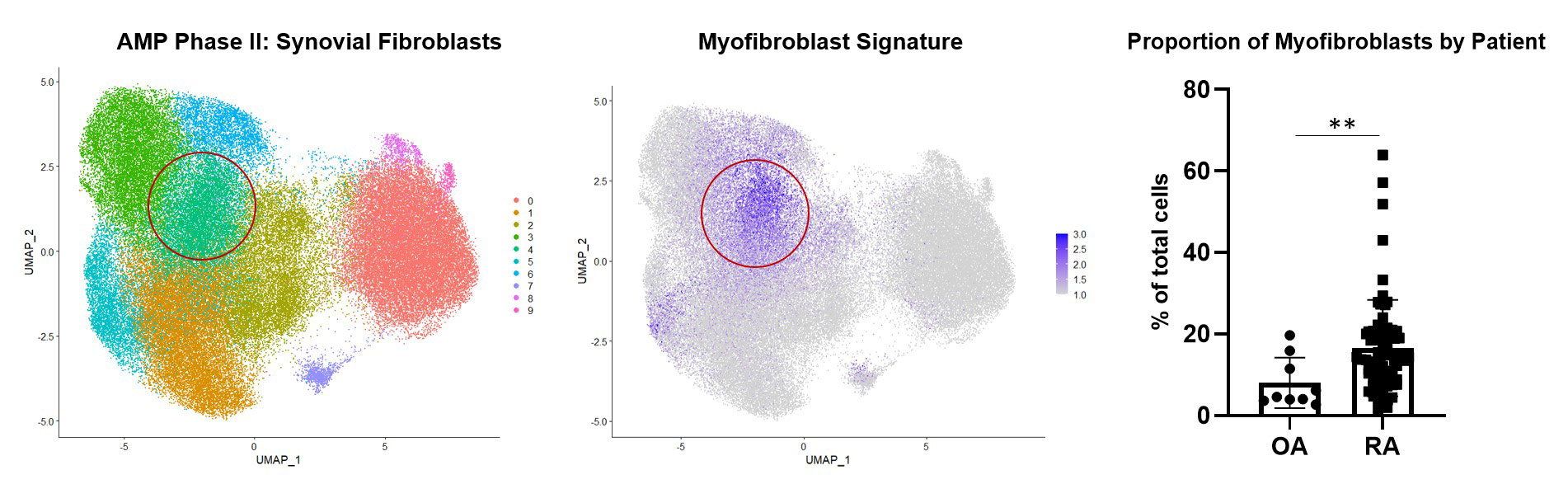
Background/Purpose: Many rheumatoid arthritis (RA) patients do not achieve sustained remission despite multiple lines of therapy. Recent studies have linked the expansion of synovial fibroblasts with treatment resistance in RA1. Using single-cell RNA-sequencing data generated by the Accelerating Medicines Partnership (AMP) Consortium, we identified the expansion of fibrotic myofibroblasts in RA distinguished by high expression of extracellular matrix proteins2. Here, we leveraged spatial transcriptomics and in vitro studies to reveal the vascular localization of myofibroblasts and identify the molecular drivers of this phenotype.

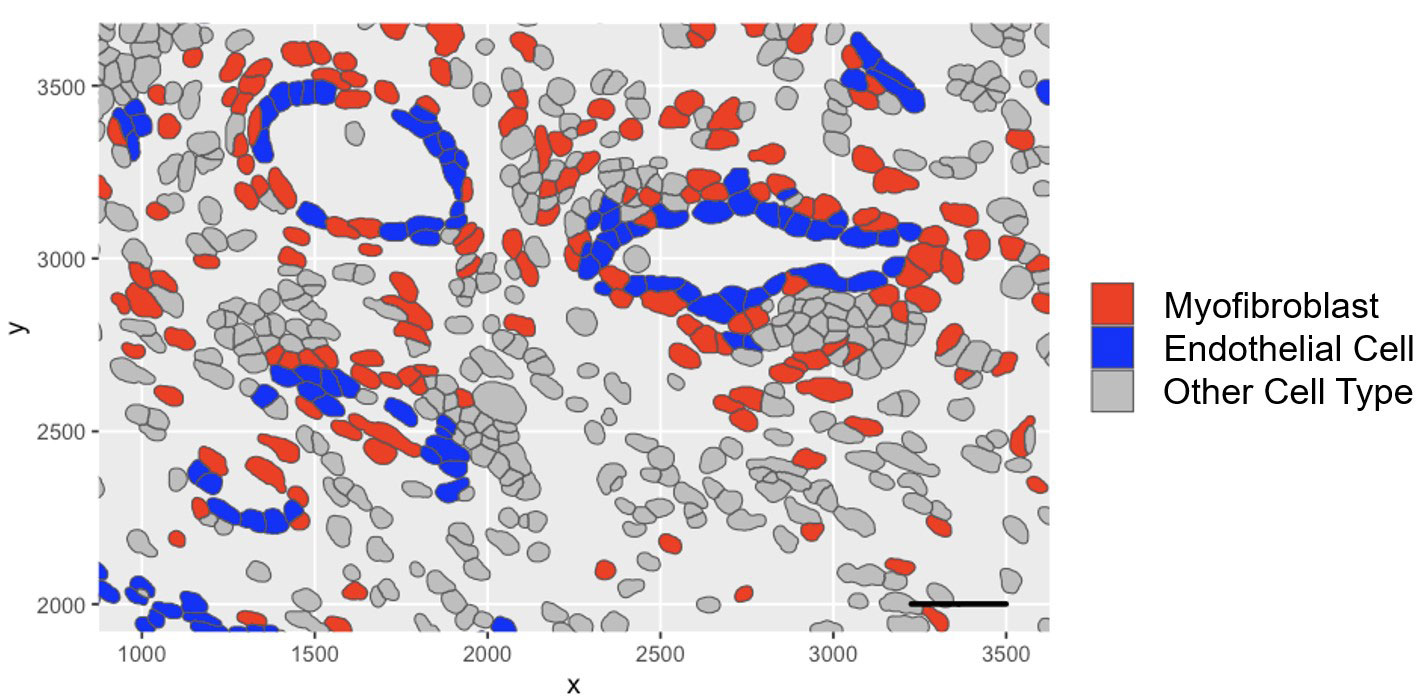
Methods: Spatial transcriptomic profiling: Synovial FFPE tissue sections (1 RA and 1 OA) were subject to cyclic in situ hybridization of 960 fluorescent mRNA probes and imaged using the CosMx™ Spatial Molecular Image (Nanostring). Cell segmentation was performed which was used to assign transcripts to cells. Subsequent clustering and visualization were performed.

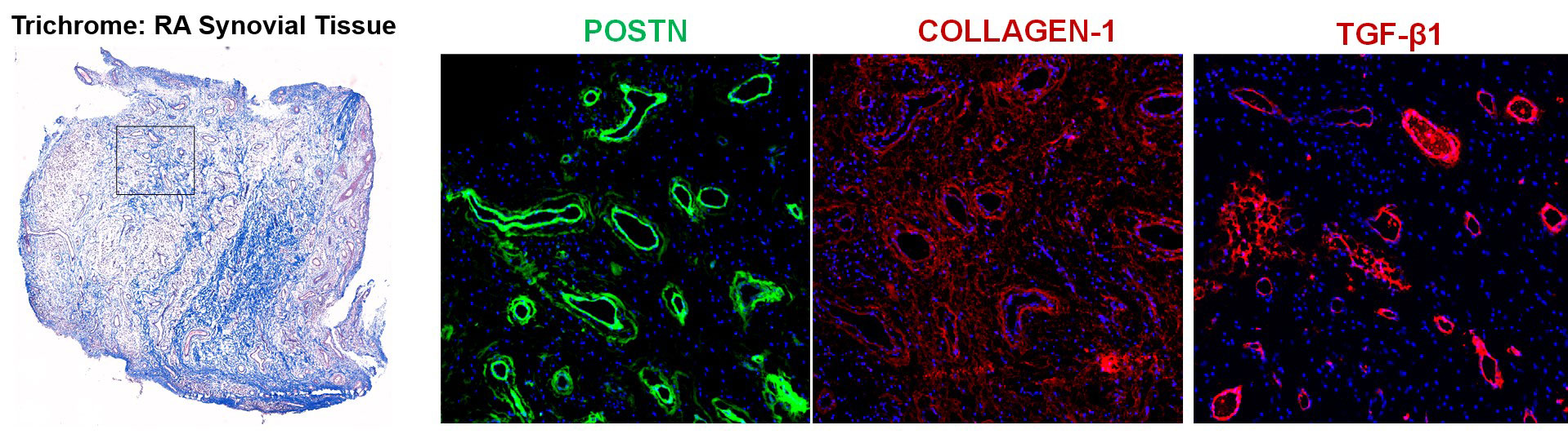
Immunofluorescent staining: RA and OA synovial tissue sections were stained with antibodies against CD31, NOTCH3, POSTN, COL1A1, p-SMAD2/3 and TGF-β followed by secondary antibody staining and image acquisition.

Cell culture: Synovial fibroblasts were cultured and stimulated with Notch ligand or co-cultured with human umbilical vein endothelial cells (HUVECs). qPCR, ELISA, western blot, and flow cytometry of stimulated cells were performed.

Results: Synovial scRNAseq data generated from 79 donors revealed the significant expansion of POSTN+ fibrotic myofibroblasts, defined by high expression extracellular matrix proteins COL1A1 and COMP, in RA. To identify the localization of myofibroblasts within RA synovia we applied a myofibroblast gene signature to spatial transcriptomic data which showed localization of myofibroblasts to the perivascular niche. Immunofluorescent staining of myofibroblast markers POSTN and COL1A1 confirmed the perivascular deposition of these proteins in RA synovia. Further scRNAseq analysis revealed a myofibroblast differentiation trajectory encompassing Notch and TGF-β signaling. Stimulation of synovial fibroblasts with Notch ligand delta-like ligand 4 and co-culture with HUVECs strongly induced the expression of POSTN. Unexpectedly, we found that Notch signaling promoted strong expression of TGF-β in vitro. Immunofluorescent staining of synovial tissue confirmed perivascular localization of NOTCH3, TGF-β and pSMAD2/3, suggesting a molecular crosstalk between Notch and TGF-β during differentiation of myofibroblasts in RA synovia.

Conclusion: Our study leveraged cutting-edge spatial transcriptomics to identify endothelial cells as mediators of synovial tissue fibrosis via cellular crosstalk involving Notch and TGF-β signaling. Further investigation into the molecular basis of myofibroblast differentiation in RA synovia will enable the identification of novel therapies targeting myofibroblasts in treatment resistant RA.

1. Rivellese, F et al. Nat Med, 19 May. 2022, doi:10.1038/s41591-022-01789-0
2. Zhang, F. et al. bioRxiv 2022.02.25.481990 (2022) doi:10.1101/2022.02.25.481990.

Supporting image 1

Figure 1: Expansion of the Myofibroblast Subset in RA

Supporting image 2

Figure 2: Spatial Transcriptomic Analysis Reveals Localization of Myofibroblasts to the Perivascular Niche

Supporting image 3

Figure 3: Extracellular Matrix Protein Deposition Occurs Perivascularly in RA Synovium


Disclosures: K. Bhamidipati, None; R. Madhu, None; S. Presti, None; Y. Kim, Nanostring Technologies; Y. Cui, Nanostring Technologies; F. Zhang, None; A. Jonsson, Amgen; A. Nathan, None; N. Millard, None; D. Rao, Janssen, Merck, Bristol-Myers Squibb, Scipher Medicine, HiFiBio, Inc., AstraZeneca, Pfizer; L. Donlin, None; J. Anolik, None; S. Raychaudhuri, Johnson & Johnson, Pfizer, Biogen, Gilead Sciences, Mestag Therapeutics; M. Brenner, GSK, 4FO Ventures, Mestag Therapeutics; I. Korsunsky, Mestag Therapeutics; K. Wei, Gilead sciences, Mestag, nanoString, 10X Genomics.

To cite this abstract in AMA style:

Bhamidipati K, Madhu R, Presti S, Kim Y, Cui Y, Zhang F, Jonsson A, Nathan A, Millard N, Rao D, Donlin L, Anolik J, Raychaudhuri S, Brenner M, Korsunsky I, Wei K. Spatial Transcriptomic Analysis Reveals Vascular Zonation of Myofibroblasts in Rheumatoid Arthritis Synovium [abstract]. Arthritis Rheumatol. 2022; 74 (suppl 9). https://acrabstracts.org/abstract/spatial-transcriptomic-analysis-reveals-vascular-zonation-of-myofibroblasts-in-rheumatoid-arthritis-synovium/. Accessed .
  • Tweet
  • Click to email a link to a friend (Opens in new window) Email
  • Click to print (Opens in new window) Print

« Back to ACR Convergence 2022

ACR Meeting Abstracts - https://acrabstracts.org/abstract/spatial-transcriptomic-analysis-reveals-vascular-zonation-of-myofibroblasts-in-rheumatoid-arthritis-synovium/

Advanced Search

Your Favorites

You can save and print a list of your favorite abstracts during your browser session by clicking the “Favorite” button at the bottom of any abstract. View your favorites »

All abstracts accepted to ACR Convergence are under media embargo once the ACR has notified presenters of their abstract’s acceptance. They may be presented at other meetings or published as manuscripts after this time but should not be discussed in non-scholarly venues or outlets. The following embargo policies are strictly enforced by the ACR.

Accepted abstracts are made available to the public online in advance of the meeting and are published in a special online supplement of our scientific journal, Arthritis & Rheumatology. Information contained in those abstracts may not be released until the abstracts appear online. In an exception to the media embargo, academic institutions, private organizations, and companies with products whose value may be influenced by information contained in an abstract may issue a press release to coincide with the availability of an ACR abstract on the ACR website. However, the ACR continues to require that information that goes beyond that contained in the abstract (e.g., discussion of the abstract done as part of editorial news coverage) is under media embargo until 10:00 AM ET on November 14, 2024. Journalists with access to embargoed information cannot release articles or editorial news coverage before this time. Editorial news coverage is considered original articles/videos developed by employed journalists to report facts, commentary, and subject matter expert quotes in a narrative form using a variety of sources (e.g., research, announcements, press releases, events, etc.).

Violation of this policy may result in the abstract being withdrawn from the meeting and other measures deemed appropriate. Authors are responsible for notifying colleagues, institutions, communications firms, and all other stakeholders related to the development or promotion of the abstract about this policy. If you have questions about the ACR abstract embargo policy, please contact ACR abstracts staff at [email protected].

Wiley

  • Online Journal
  • Privacy Policy
  • Permissions Policies
  • Cookie Preferences

© Copyright 2025 American College of Rheumatology