Session Information
Date: Tuesday, November 7, 2017
Title: Osteoarthritis – Clinical Aspects Poster II: Observational and Epidemiological Studies
Session Type: ACR Poster Session C
Session Time: 9:00AM-11:00AM
Background/Purpose: The knee adduction moment (KAM) estimates medial knee load during gait, with high KAM fueling worsening OA. Understanding how foot mechanics contribute to KAM would inform the development of more effective footwear and insoles to prevent worsening knee OA. During the initial and early stance phases, a neutrally aligned foot with adequate pronation could dissipate impact loads, reduce varus thrust, and minimize KAM as it rises to an initial peak. Yet, excessive pronation in late stance redistributes load medially, possibly increasing KAM as it reaches a second peak. In persons with medial knee OA, we assessed: 1) the relation of foot pronation during the initial and early stance phases to 1st peak KAM; and 2) the relation of foot pronation during late stance to 2nd peak KAM. Given possible effects of both diminished and excessive pronation, we assessed both linear and u-shaped trends.
Methods: The Effectiveness of Shoes and Insoles on Loading at the Knee (SILK) Study included adults aged 45-70 yrs with medial knee OA. Plantar pressure profiles of the index limb were acquired during 5 trials of self-paced barefoot walking using an Emed-X platform. From these, we measured foot pronation during the initial, early, and late stance phases using the Pronation-Supination Index (PSI) as described by Motooka et al., where lower PSI values indicate greater pronation (retest ICC 0.46-0.90). Concurrently, a Qualisys motion capture system with AMTI force plates measured 1st and 2nd peak KAM in Newton-meters per kg of body weight (retest ICC= 0.86). In tertiles of increasing pronation (decreasing PSI), generalized linear models estimated mean 1st and 2nd peak KAM while adjusting for age, sex, body mass, and walking velocity. Tests for linear and u-shaped trend identified dose-response relationships.
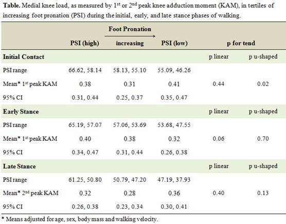
Results: In 70 SILK participants (mean age 60.3 ± 9.6 yrs, body mass 87.3 ± 18.5 kg, walking velocity 1.12 ± 0.23 m/sec, 61.4% female), mean 1st and 2nd peak KAM was 0.36 and 0.32 Nm/kg, respectively. At initial contact, feet with diminished (PSI ≥ 58.14) or increased (PSI ≤ 55.09) pronation in comparison to the middle PSI tertile (PSI 55.10, 58.13) had increased 1st peak KAM (mean difference = 0.07 and 0.10 Nm/kg, respectively), resulting in a significant u-shaped trend (p for trend = 0.02). During early stance, 1st peak KAM was greatest among feet with the least pronation and declined linearly across tertiles of increasing pronation (p for trend = 0.06). There was no association between foot pronation in late stance and 2nd peak KAM (see table).
Conclusion: In adults with medial knee OA, feet with diminished or excessive pronation at initial contact, and feet with reduced pronation during the early stance phase of walking, experience increased peak medial knee loads. These findings could inform efforts to refine more effective footwear or insole interventions to minimize loads for persons with medial knee OA.
To cite this abstract in AMA style:
Gross KD, Jones R, Felson DT, Chong S, Hillstrom HJ. Relation of Foot Pronation to Medial Knee Load in Persons with Medial Knee Osteoarthritis [abstract]. Arthritis Rheumatol. 2017; 69 (suppl 10). https://acrabstracts.org/abstract/relation-of-foot-pronation-to-medial-knee-load-in-persons-with-medial-knee-osteoarthritis/. Accessed .« Back to 2017 ACR/ARHP Annual Meeting
ACR Meeting Abstracts - https://acrabstracts.org/abstract/relation-of-foot-pronation-to-medial-knee-load-in-persons-with-medial-knee-osteoarthritis/