Session Information
Session Type: ACR Poster Session A
Session Time: 9:00AM-11:00AM
Background/Purpose:
Foot pronation can lead to lateral patellofemoral (PF) maltracking with reduced tibiofemoral (TF) varum, inciting speculation that the highly pronated foot reported in knees with osteoarthritis (OA) could increase risk of worsening lateral PF joint damage, while reducing risk of worsening medial TF damage. Yet, longitudinal data is lacking and dynamic foot pronation measured during the early, mid, and terminal phases of walking may differ from measures in static standing. Our purpose was to explore the relation of foot pronation measured during each phase of walking to the 2-year risk of worsening lateral PF and medial TF cartilage damage. Pronation effects on PF tracking and knee varus moment tend to be maximal during the early stance phase.Methods:
The Multicenter Osteoarthritis Study (MOST) includes adults aged 50-79 years with or at risk of knee OA. At the 60 month exam, high-resolution plantar pressure profiles were acquired during 5 trials of self-paced walking. From these, we measured foot pronation during the early, mid, and terminal stance phases using the Pronation-Supination Index (PSI), as described by Motooka, et al. (same-day retest ICC 0.46 to 0.94), where lower values indicate greater pronation. From 1.0 T MRIs, readers scored (0-6) one knee per subject for cartilage damage in each sub-region of the lateral PF (2 sub-regions) and medial TF (5 sub-regions) compartments using WORMS (weighted kappa > 0.63). Among sub-regions with submaximal scores at 60 months, worsening damage was any increase in WORMS score at 84 months. Separate logistic regression models estimated the relative odds of worsening lateral PF and medial TF cartilage damage within quintiles of increasing foot pronation compared to feet with the least pronation. Adjustments were made for age, sex, and BMI. GEE accounted for non-independent knee sub-regions.Results:
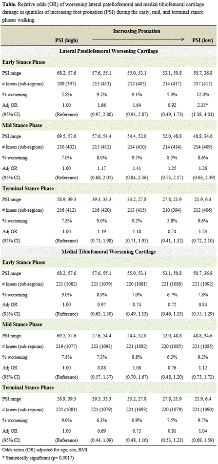
1067 and 1104 subjects (mean age 66.8 ± 7.6 years, BMI 29.6 ± 4.8 kg/m2, 60.7% female) contributed to the analysis of worsening lateral PF and medial TF cartilage damage, respectively. Compared to feet with the least pronation, odds of worsening lateral PF damage were 2.35 (95% CI: 1.38, 4.01) times greater in limbs with the greatest foot pronation during early stance (p < 0.002). A non-significant trend suggested a possible relation between greater early phase foot pronation and reduced odds of medial TF worsening. There was no association between foot pronation during other phases of walking and risk of worsening knee damage (Table).Conclusion:
In adults with or at-risk of knee OA, feet with the greatest foot pronation during the early stance phase of walking are at increased risk of worsening knee cartilage damage in the lateral PF compartment. These findings could inform efforts to refine preventative footwear interventions for compartment-specific knee OA.
Disclosure: K. D. Gross, None; H. J. Hillstrom, None; C. Brown, None; R. Jones, None; J. Stefanik, None; M. C. Nevitt, None; C. E. Lewis, None; J. Torner, None; D. T. Felson, zimmer knee creations, 5.
To cite this abstract in AMA style:
Gross KD, Hillstrom HJ, Brown C, Jones R, Stefanik J, Nevitt MC, Lewis CE, Torner J, Felson DT. Relation of Foot Pronation during Walking to Risk of Worsening Lateral Patellofemoral and Medial Tibiofemoral Cartilage Damage [abstract]. Arthritis Rheumatol. 2016; 68 (suppl 10). https://acrabstracts.org/abstract/relation-of-foot-pronation-during-walking-to-risk-of-worsening-lateral-patellofemoral-and-medial-tibiofemoral-cartilage-damage/. Accessed .« Back to 2016 ACR/ARHP Annual Meeting
ACR Meeting Abstracts - https://acrabstracts.org/abstract/relation-of-foot-pronation-during-walking-to-risk-of-worsening-lateral-patellofemoral-and-medial-tibiofemoral-cartilage-damage/

