Session Information
Date: Monday, October 27, 2025
Title: (1191–1220) Muscle Biology, Myositis & Myopathies – Basic & Clinical Science Poster II
Session Type: Poster Session B
Session Time: 10:30AM-12:30PM
Background/Purpose: Idiopathic inflammatory myopathies (IIM) involve muscle inflammation. MicroRNAs (miRNAs), such as miR-133a-3p and miR-1-3p, play a key role in gene regulation and muscle repair. Their dysregulated expression contributes to IIM pathogenesis, making them potential biomarkers and therapeutic targets. This study evaluates the expression of miR-133a-3p and miR-1-3p in IIM vs. healthy controls (HC), and the association with muscle involvement and disease-related features, and the impact of initial immunosuppressive therapy.
Methods: 60 patients with IIM were included: 19 males; mean (SD) age 59.1 (12.9) years; disease duration 2.5 (3.5) years; 20 dermatomyositis (DM)/ 25 polymyositis (PM), both without statin history/ 15 statin-induced immune-mediated necrotizing myopathies (IMNM). Follow-up at 6 months of immunosuppressive therapy included 13 patients (5 DM, 5 PM, 3 IMNM) with early disease (disease duration 0.9 (1.3) years). Furthermore, 33 age-/sex-matched HC were included (10 males, age 58.3 (13.4)). Body composition was examined using bioelectric impedance analysis (BIA-2000-M). Total RNA was extracted from plasma with miRNeasy Serum/Plasma Advanced kit (Qiagen). Quantitative PCR was performed using 6 miRCURY LNA miRNA PCR assays (Qiagen) with miR-103-3p, miR-191-5p, and let-7a-5p as endogenous controls. miRNA concentration analysis was performed using GenEx software (Multid Analysis AB). Data are presented as mean (SEM).
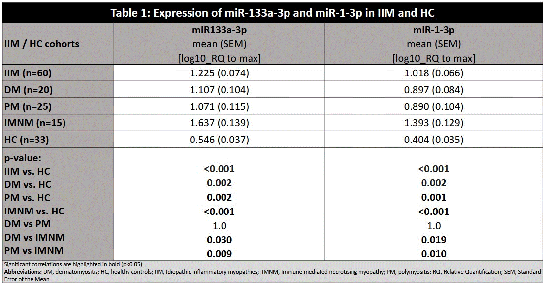
Results: The expression of miR-133a-3p and miR-1-3p was significantly higher in IIM vs. HC, in all IIM subsets vs. HC, and particularly in IMNM vs. DM and PM (Table 1) and correlated with increased muscle damage markers (CK, LD, myoglobin), decreased muscle strength (MMT8), and shorter disease duration (Table 2). After 6 months of immunosuppressive therapy, miR-133a-3p and miR-1-3p levels significantly decreased, alongside disease activity (MYOACT) and muscle damage markers, while muscle strength (MMT-8) improved (Table 3). A greater miR-133a-3p decrease correlated with a larger decrease in CK (p=0.003; r=0.780) and LD (p=0.030; r=0.623), and strength improvement (p=0.043; r=0.591). Similarly, larger miR-1-3p reduction correlated with greater CK decrease (p=0.010; r=0.705). Higher baseline miR-133a-3p and/or miR-1-3p predicted greater muscle strength improvement (p=0.043; r=0.591) and larger reductions in CK (p=0.007; r=0.727 and p=0.017; r=0.671), LD (p=0.010; r=0.707 and p=0.075; r=0.532), and myoglobin (p=0.059; r=0.584, respectively).
Conclusion: The expression of miR-133a-3p and miR-1-3p were significantly higher in IIM vs. HC, in all IIM subsets vs. HC, particularly in IMNM than in DM and PM, reflecting greater muscle damage. The levels were highest in early disease with high activity and declined after 6 months of immunosuppressive therapy, mirroring reduced muscle damage markers and improved strength. Our study demonstrated both miR-133a-3p and miR-1-3p as a potential biomarker of disease activity and muscle damage in IIM, and a potential predictor of initial treatment response in IIM.Acknowledgment: MH CR 023728, NU21-01-00146, NU21-05-00322, BBMRI.cz-LM2023033, SVV 260638
To cite this abstract in AMA style:
Svobodová K, Oreská S, Vernerová L, Dlouhá D, Wunsch H, Pavelka K, Vencovsky J, Šenolt L, Vrablik M, Hubáček J, Tomcik M. Plasma Levels of miR-133a-3p and miR-1-3p as Potential Biomarkers of Muscle Involvement and Initial Treatment Response in Myositis [abstract]. Arthritis Rheumatol. 2025; 77 (suppl 9). https://acrabstracts.org/abstract/plasma-levels-of-mir-133a-3p-and-mir-1-3p-as-potential-biomarkers-of-muscle-involvement-and-initial-treatment-response-in-myositis/. Accessed .« Back to ACR Convergence 2025
ACR Meeting Abstracts - https://acrabstracts.org/abstract/plasma-levels-of-mir-133a-3p-and-mir-1-3p-as-potential-biomarkers-of-muscle-involvement-and-initial-treatment-response-in-myositis/


.gif)
.gif)