ACR Meeting Abstracts

ACR Meeting Abstracts

  • Meetings
    • ACR Convergence 2024
    • ACR Convergence 2023
    • 2023 ACR/ARP PRSYM
    • ACR Convergence 2022
    • ACR Convergence 2021
    • ACR Convergence 2020
    • 2020 ACR/ARP PRSYM
    • 2019 ACR/ARP Annual Meeting
    • 2018-2009 Meetings
    • Download Abstracts
  • Keyword Index
  • Advanced Search
  • Your Favorites
    • Favorites
    • Login
    • View and print all favorites
    • Clear all your favorites
  • ACR Meetings

Abstract Number: 0662

Partial Correlations Network Models Show Th1, Th2 and Th17 Responses to Be Interlinked in Dermal Pathogenesis of Cutaneous Lupus Erythematous

Felix Chin1, Thomas Vazquez2, Josh Dan3, DeAnna Diaz4, Grant Sprow5, Jay Patel6, Nilesh Kodali7, Rui Feng8 and Victoria Werth9, 1University of Pennsylvania Perelman School of Medicine, Philadelphia, PA, 2FIU Wertheim College of Medicine, Virginia Beach, VA, 3Philadelphia VAMC, Philadelphia, PA, USA and Department of Dermatology, Perelman School of Medicine at the University of Pennsylvania, Philadelphia, 4Philadelphia College of Medicine, Philadelphia, PA, 5Albert Einstein College of Medicine, Philadelphia, PA, 6Philadelphia VAMC, Philadelphia, PA, USA and Department of Dermatology, Perelman School of Medicine at the University of Pennsylvania, Philadelphia, PN, 7New Jersey Medical School, Coppell, TX, 8University of Pennsylvania, Philadelphia, 9University of Pennsylvania and Corporal Michael J. Crescenz VA Medical Center, Philadelphia, PA

Meeting: ACR Convergence 2022

Keywords: autoimmune diseases, Dermatology, immunology, lupus-like disease, T Cell

  • Tweet
  • Email
  • Print
Session Information

Date: Sunday, November 13, 2022

Title: SLE – Etiology and Pathogenesis Poster

Session Type: Poster Session B

Session Time: 9:00AM-10:30AM

Background/Purpose: The immunopathogenesis of cutaneous lupus erythematous (CLE) is highly diverse and involves activity of many different cell types and pathways. This heterogeneity is believed to be largely responsible for unpredictable treatment response between patients. Here we used a correlation network model approach to map the relationships between different cell types and pathway proteins in CLE skin biopsies.

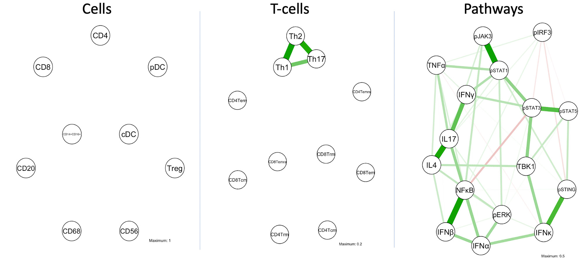
Methods: Here we profiled 44 CLE biopsies using multiplexed imaging mass cytometry to characterize the cell and pathway composition of CLE infiltrate. Biopsies were stained with two different panels of 37-metal conjugated antibodies that served as markers for different cell types, cytokines and pathway proteins. The relationships among cell and cytokine immune markers were modeled using Gaussian graphical model. The values were log-transformed and standardized prior to the analysis. The GGM algorithm identifies and displays only significant correlations between markers while leaving out non-significant ones. The algorithm then graphs the markers as a network and spatially arranges them by correlation strength so that the strongest associated markers are within closest proximity. This spatial orientation allows for distinction between direct and indirect associations of cell types or pathway proteins.

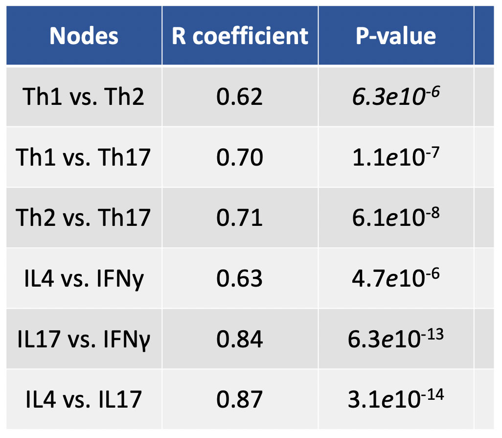
Results: Of our network consisting of 11 T-cell subtypes, GGM identified Th1, Th2 and Th17 to be inter-related with each other, forming a triangle. No other positive findings were found between any other cells, including our main cell network consisting of 11 common immune cell types. Of our cytokine/pathway network consisting of 16 different markers, GGM identified IL4, IL17 and IFN???? to be among the strongest related markers in the network. Correlation testing confirmed remarkably strong associations between IL4, IL17 and IFN???? (all 3 pairs were p< 1×10-13) as well as significant correlations (p< 1×10⁻⁶) amongst all possible permutations of the aforementioned GGM-significant markers. Furthermore, heatmap visualization of T-cell K-means clustering was also performed and revealed the majority of patients (n=27) to be Tem dominant (83% CD4 and 76% CD8), with a smaller subgroup (n=14) to being Tcm dominant (57% CD4 and 50% CD8).

Conclusion: These findings suggest that Th1, Th2 and Th17 co-occur together in the pathogenesis of CLE. Th1 and Th2 responses have traditionally been thought of as separate and antagonistic to each other. Our study challenges this dualism. In treating CLE patients with increased T-cell activity, it may be necessary to tackle both Th1 and Th2 along with Th17 responses together.

Supporting image 1

Fig 1. GGM networks of cells, T-cell subsets and pathway proteins. Lines indicate significant correlations between nodes. Green lines indicate positive association while red lines indicate a negative association. Line color intensity and spatial proximity between nodes is a reflection of correlation strength.

Supporting image 2

Table 1. Spearman’s R correlation coefficient strengths between all possible pairs of cells or cytokines involved in Th1, Th2 and Th17 response.

Supporting image 3

Fig 2. Linear relationship between IL4 and IL17 levels across 44 patients studied.


Disclosures: F. Chin, None; T. Vazquez, None; J. Dan, None; D. Diaz, None; G. Sprow, None; J. Patel, None; N. Kodali, None; R. Feng, None; V. Werth, AbbVie/Abbott, Amgen, AstraZeneca, Bristol Myers Squibb (BMS), Celgene, Eli Lilly, Genentech, GlaxoSmithKline (GSK), Janssen, Merck/MSD, Gilead, Novartis, Pfizer, Rome Pharmaceuticals, Horizon Therapeutics, Regeneron, argenx, CSL Behring, AnaptysBio, Biogen, Corbus, EMD Serono, Galderma, Nektar, Octapharma, Principia, Resolve, Sanofi, Syntimmune, Viela.

To cite this abstract in AMA style:

Chin F, Vazquez T, Dan J, Diaz D, Sprow G, Patel J, Kodali N, Feng R, Werth V. Partial Correlations Network Models Show Th1, Th2 and Th17 Responses to Be Interlinked in Dermal Pathogenesis of Cutaneous Lupus Erythematous [abstract]. Arthritis Rheumatol. 2022; 74 (suppl 9). https://acrabstracts.org/abstract/partial-correlations-network-models-show-th1-th2-and-th17-responses-to-be-interlinked-in-dermal-pathogenesis-of-cutaneous-lupus-erythematous/. Accessed .
  • Tweet
  • Email
  • Print

« Back to ACR Convergence 2022

ACR Meeting Abstracts - https://acrabstracts.org/abstract/partial-correlations-network-models-show-th1-th2-and-th17-responses-to-be-interlinked-in-dermal-pathogenesis-of-cutaneous-lupus-erythematous/

Advanced Search

Your Favorites

You can save and print a list of your favorite abstracts during your browser session by clicking the “Favorite” button at the bottom of any abstract. View your favorites »

All abstracts accepted to ACR Convergence are under media embargo once the ACR has notified presenters of their abstract’s acceptance. They may be presented at other meetings or published as manuscripts after this time but should not be discussed in non-scholarly venues or outlets. The following embargo policies are strictly enforced by the ACR.

Accepted abstracts are made available to the public online in advance of the meeting and are published in a special online supplement of our scientific journal, Arthritis & Rheumatology. Information contained in those abstracts may not be released until the abstracts appear online. In an exception to the media embargo, academic institutions, private organizations, and companies with products whose value may be influenced by information contained in an abstract may issue a press release to coincide with the availability of an ACR abstract on the ACR website. However, the ACR continues to require that information that goes beyond that contained in the abstract (e.g., discussion of the abstract done as part of editorial news coverage) is under media embargo until 10:00 AM ET on November 14, 2024. Journalists with access to embargoed information cannot release articles or editorial news coverage before this time. Editorial news coverage is considered original articles/videos developed by employed journalists to report facts, commentary, and subject matter expert quotes in a narrative form using a variety of sources (e.g., research, announcements, press releases, events, etc.).

Violation of this policy may result in the abstract being withdrawn from the meeting and other measures deemed appropriate. Authors are responsible for notifying colleagues, institutions, communications firms, and all other stakeholders related to the development or promotion of the abstract about this policy. If you have questions about the ACR abstract embargo policy, please contact ACR abstracts staff at [email protected].

Wiley

  • Online Journal
  • Privacy Policy
  • Permissions Policies
  • Cookie Preferences

© Copyright 2025 American College of Rheumatology