Session Information
Session Type: Poster Session D
Session Time: 9:00AM-11:00AM
Background/Purpose: Morphea is an autoimmune disorder causing sclerosis and inflammation of the skin and subcutaneous tissue. Facial morphea can cause substantial disfigurement and negative impact on quality of life. Existing clinical measures such as the LoSCAT likely fail to capture the severity of facial morphea due to their body site driven approach and difficulty in quantifying tissue loss making it difficult to assess severity and response to treatment. 3D stereophotogrammetry is a non-invasive, readily available modality that has been used measure facial asymmetry in disorders such as cleft palate. The aim of this pilot study was to investigate the feasibility of use of this modality in identifying asymmetry in facial morphea, and to compare 3D stereophotogrammetry to clinical assessment.
Methods: 23 participants with facial morphea were recruited from the Morphea in Adults and Children Cohort at UT Southwestern Medical Center. They were evaluated by a single dermatologist (HJ), at which time they were assigned a LoSCAT score, a clinical morphea subtype, and completed validated quality of life questionnaires. Participants then underwent 3D stereophotogrammetry. Facial asymmetry (mm) was determined by calculating the distance between layers of stereolithography images using an in-house written mathematical model in MATLAB, thus quantifying the amount of atrophy (Figure 1 gives an example of a 3D image and heat map generated from this technique). In order to analyze asymmetry in specific anatomical regions of interest, established anatomical landmarks were used to segment facial subunits of interest, and both mean and maximum 5% values were calculated in order to account for dilution.
Results: The majority of patients were children (70%), female (74%), Caucasian (61%), and had linear morphea (en coup de sabre) (52%). Table 1 provides demographic and clinical information of participants. Based on a previously published standard of 1.2mm to define pathologic asymmetry, 22 of 23 patients showed pathologic asymmetry in every facial subunit (Table 2 provides asymmetry values). In order of greatest to least asymmetry, the facial subunits were ranked as follows: forehead, cheek, nose, then mouth. While LoSDI scores did not correlate with asymmetry values, PGA-D values correlated significantly to mean mouth (p=0.0021) and cheek asymmetry (p=0.04). Quality of life measures also showed correlation with asymmetry in the lower face (p=0.013).
Conclusion: Results of this pilot study indicate that the majority of facial morphea lesions produce asymmetry that can be quantified using 3D stereophotogrammetry. Interestingly, we found that both physicians and patients attributed greater severity to lesions affecting the lower part of the face while actual percent asymmetry was greatest in the forehead, indicating that percent asymmetry alone does not drive impact on patients. Taken together, these results confirm that the LoSCAT alone is an inadequate measure of facial morphea and the use of 3D facial imaging maybe a valuable addition to evaluation. Further studies using this modality are needed to determine clinically important difference in facial asymmetry and sensitivity to change.
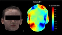 Figure 1 shows a 3D image and heat map created by the 3D stereophotogrammetry technique, which demonstrates pathologic asymmetry in a morphea patient.
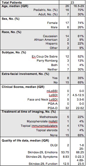 Table 1 provides demographic and clinical information of participants.
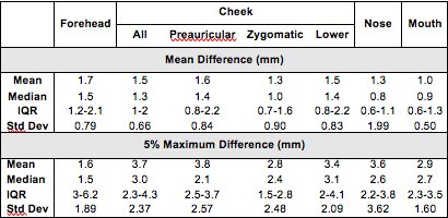 Table 2 provides asymmetry values in millimeters of study patients as determined through 3D stereophotogrammetry.
To cite this abstract in AMA style:
Abbas L, Day J, Jacobe H. Measuring Asymmetry in Facial Morphea via 3D Stereophotogrammetry [abstract]. Arthritis Rheumatol. 2020; 72 (suppl 10). https://acrabstracts.org/abstract/measuring-asymmetry-in-facial-morphea-via-3d-stereophotogrammetry/. Accessed .« Back to ACR Convergence 2020
ACR Meeting Abstracts - https://acrabstracts.org/abstract/measuring-asymmetry-in-facial-morphea-via-3d-stereophotogrammetry/