Session Information
Date: Saturday, November 16, 2024
Title: Metabolic & Crystal Arthropathies – Basic & Clinical Science Poster I
Session Type: Poster Session A
Session Time: 10:30AM-12:30PM
Background/Purpose: Gout, due to the presence of monosodium urate crystals (MSU) and calcium pyrophosphate (CPP) crystal deposition disease are both responsible for recurrent inflammation flares. Both crystals stimulate interleukin-1β production by resident macrophages. Macrophages can interact and phagocytosis both MSU and CPP crystals.
Objective: to describe the intracellullar fates of MSU and CPP crystals in macrophages
Methods: Synthetic pyrogen-free MSU and CPP crytals were used to stimulate different type of mouse (RAW 264.7 cell line, bone marrow-derived macrophages (BMDM)) and human (THP-1 cell line and peripheral blood mononuclear cells (PBMC)) macrophages. To assess crystal phagocytosis, macrophages were stimulated by MSU and CPP crystals engrafted with fluorescent organic nanoparticles (FON) and recorded during 5 days using Incucyte Live-cell Analysis instrument (Sartorius). Intracellular outcomes were assessed by 18-hours recording using apoptome Zeiss microscopy. Images analysis was performed by ImageJ software.
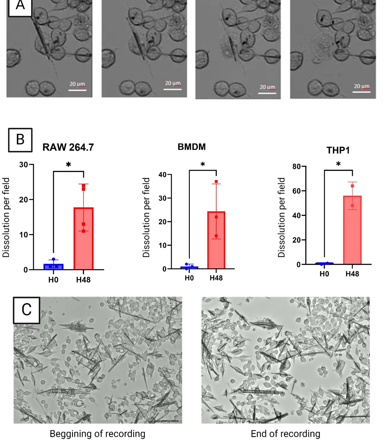
Results: All type of macrophages including RAW 264.7, BMDM, THP-1 and PBMC, rapidly phagocytosed both MSU and CPP crystals. Crystal phagocytosis occurred within the first minutes and all crystals were phagocytosed within one hour. After phagocytosed, crystals remained mostly in the cytosol. Interestingly, in few RAW 264.7 macrophages and PBMC, phagocytosed MSU crystals were expulsed into the cytosol of another macrophage. MSU crystals were dissolved after phagocytosis while CPP crystals remained undissolved even after 7 days of stimulation. MSU crystals dissolution occurred only 48 hours after phagocytosis. No dissolution was observed in the first 48 hours while all MSU crystals were dissolved after 7 days of stimulation. Macrophages displayed different capacity to dissolve MSU crystals: after 48 hours of stimulation, 17.5% of MSU crystals (in one recording field) were dissolved in RAW 264.7 cells vs 24.3% and 56.0% for BMDM and THP-1, respectively. Interestingly, we observed that dissolution of MSU crystals was followed by cell death and burst in RAW 264.7 macrophages and PBMC. THP-1 cells also dissolved MSU crystals but without burst nor death. Interestingly, treatment with the inhibitor of the V-ATPase pump, bafilomycin A1, completely blocked the dissolution of MSU crystals without affecting crystal phagocytosis.
Conclusion: Our result suggest that dissolution of MSU crystals is a late cellular response involving cell lysosomes and acidification. Furthers studies are ongoing to decipher the underlying mechanisms.
To cite this abstract in AMA style:
Leroy C, Pham N, Brial F, Kischkel B, Jayat g, Ishow E, Combes C, Joosten L, Latourte A, Richette P, Ea H. Macrophage Intracellular Fates of Monosodium Urate and Calcium Pyrophosphate Crystals: Phagocytosis, Exchanged/expulsion and Dissolution of Crystals [abstract]. Arthritis Rheumatol. 2024; 76 (suppl 9). https://acrabstracts.org/abstract/macrophage-intracellular-fates-of-monosodium-urate-and-calcium-pyrophosphate-crystals-phagocytosis-exchanged-expulsion-and-dissolution-of-crystals/. Accessed .« Back to ACR Convergence 2024
ACR Meeting Abstracts - https://acrabstracts.org/abstract/macrophage-intracellular-fates-of-monosodium-urate-and-calcium-pyrophosphate-crystals-phagocytosis-exchanged-expulsion-and-dissolution-of-crystals/

