ACR Meeting Abstracts

ACR Meeting Abstracts

  • Meetings
    • ACR Convergence 2024
    • ACR Convergence 2023
    • 2023 ACR/ARP PRSYM
    • ACR Convergence 2022
    • ACR Convergence 2021
    • ACR Convergence 2020
    • 2020 ACR/ARP PRSYM
    • 2019 ACR/ARP Annual Meeting
    • 2018-2009 Meetings
    • Download Abstracts
  • Keyword Index
  • Advanced Search
  • Your Favorites
    • Favorites
    • Login
    • View and print all favorites
    • Clear all your favorites
  • ACR Meetings

Abstract Number: 0261

Machine Learning and Computational Pathology Can Reveal Synovial Pathotypes in Mice and Humans

Richard Bell1, Matthew Brendel2, Justin Xiang3, AMP RA/SLE Consortium1, Edward DiCarlo1, Jennifer Anolik4, Laura Donlin1, Dana Orange5, Edward Schwarz4, Lionel Ivashkiv1 and Fei Wang2, 1Hospital for Special Surgery, New York, NY, 2Weil Cornell Medical College, New York, NY, 3Horace Grant High School, New York, NY, 4University of Rochester Medical Center, Rochester, NY, 5The Rockefeller University, New York, NY

Meeting: ACR Convergence 2022

Keywords: autoimmune diseases, immunology, rheumatoid arthritis, Synovitis

  • Tweet
  • Email
  • Print
Session Information

Date: Saturday, November 12, 2022

Title: RA – Diagnosis, Manifestations, and Outcomes Poster I

Session Type: Poster Session A

Session Time: 1:00PM-3:00PM

Background/Purpose: Synovial pathotyping is a critical step in understanding the etiology of inflammatory arthritis as different pathotypes have differential response to therapy. Determining these pathotypes relies on immunohistochemistry of lymphoid, myeloid and stromal cells with expert grading of histologic sections, which can be time consuming and costly. Here, we developed an automated segmentation and cell type classification pipeline to efficiently phenotype a murine inflammatory arthritis model; and demonstrate the feasibility of this analysis in human synovial biopsies.

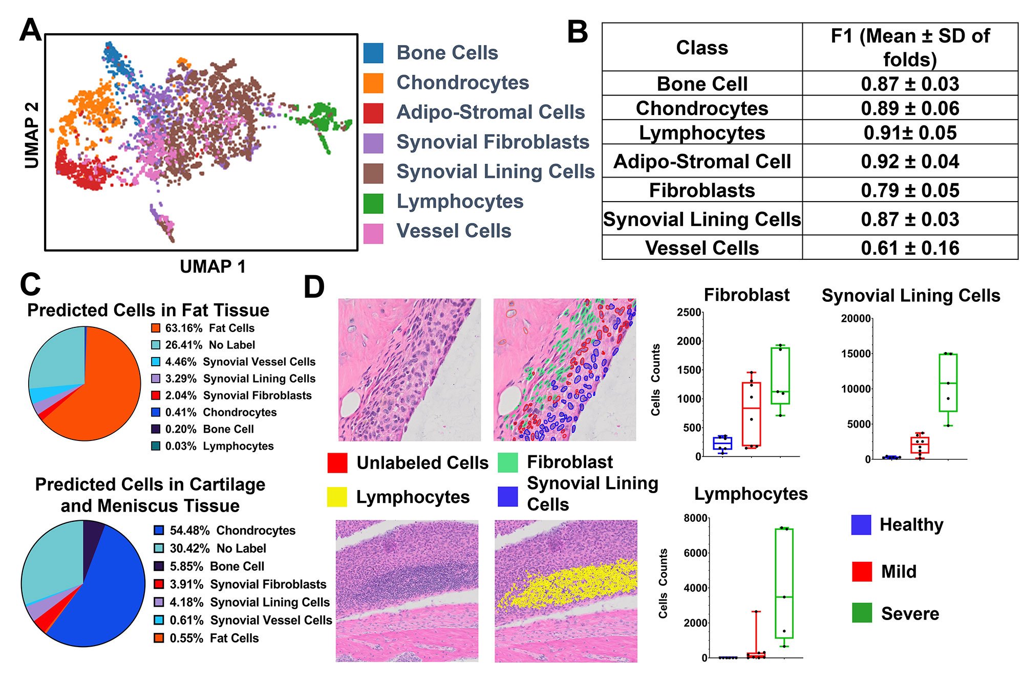
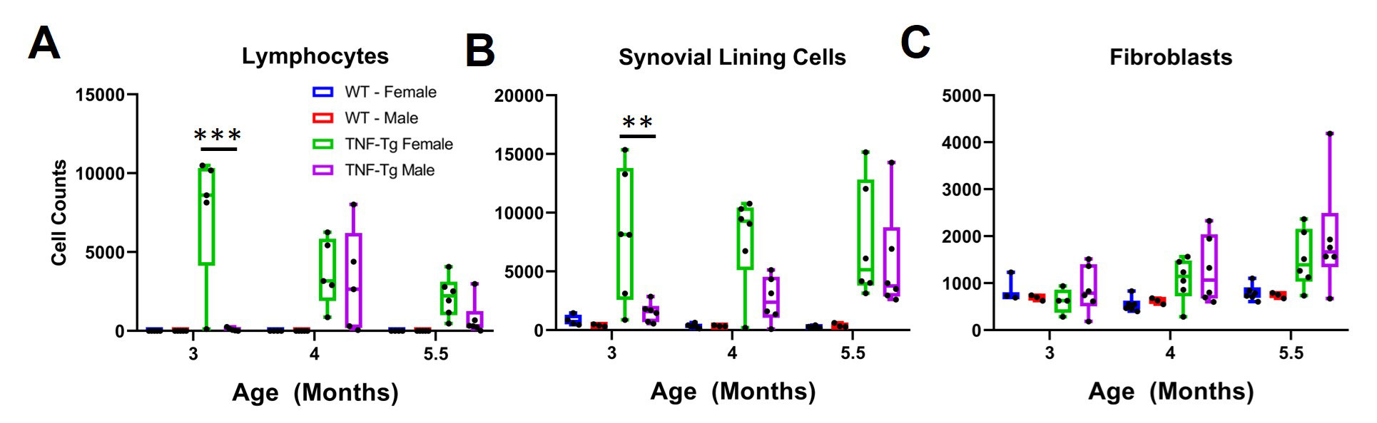
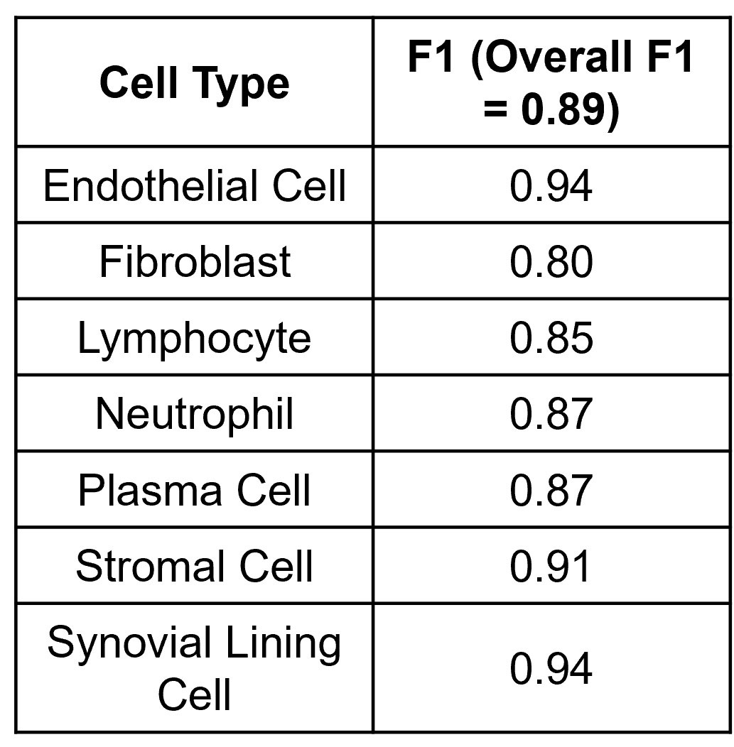
Methods: The synovial tissue, cartilage, bone, and fatty tissue from H&E sections of healthy (n=6), mild (n=8) and severely diseased (n=5) TNF transgenic mice that develop arthritis (Training and Testing Slide) was segmented using a knee tissue segmentation model previously developed in our lab1. We then annotated bone cells (n=312), vessel cells (n=378), adipo-stromal cells (n=506), fibroblasts (n=749), chondrocytes (n=625), lymphocytes (n=467), and all other synovial lining cells (n=1675). Nuclei were segmented and cellular image features were extracted in ImageJ/Python. A stratified 5-fold cross validation strategy was used to train a Gradient Boosted Decision Tree to classify cell types and F1 scores were calculated. Another set of 68 TNF-Tg slides2 (External Validation) describing the model’s sexual dimorphism were subjected to the tissue segmentation, nuclear segmentation, and cell feature extraction and cell types from the synovial tissue were inferred to validate the ability to phenotype the synovium. Also, five human synovial biopsies H&E sections from the Accelerating Medicine Partnership – Rheumatoid Arthritis consortium were annotated for 7 cell types: endothelial cell (n=117), fibroblasts (n=61), lymphocytes (n=71), neutrophils (n=93), plasma cells (n=77), stromal cells (n=57) and synovial lining cells (n=26). The same image analysis and machine learning pipeline was performed.

Results: UMAP demonstrated good separation of TNF-Tg cells and our classification model performed well (Fig 1A, B). We inferred the cell types of the remaining cells on the training and testing slides and demonstrated the tissue specificity of adipo-stromal cells and chondrocytes as the major cell type within their respective tissue (Fig1 C). When stratified by disease severity, fibroblasts, lining cells and lymphocytes followed the expected distribution (Fig 1D). In the external validation set, we also observed the expected increase in synovial lymphocytes and stromal cells in TNF-Tg female mice compared to males at earlier timepoints, but interestingly not in the fibroblast compartment, which suggested a differential etiology (Fig 2A-C). Our pipeline is also able to confidently classify 7 cell types in human RA synovial biopsies, suggesting that we might be able to incorporate our process into other synovial pathotyping pipelines (Fig 3).

Conclusion: We developed a computational pathology cell classification pipeline that confidently identifies cell types in murine and human inflammatory arthritis H&E stained tissue sections. This will allow for efficient pathotyping of diseased tissues to elucidate varying etiologies.

Supporting image 1

Figure 1. Cell type classification model successfully identifies important cell types in inflammatory arthritis. Utilizing (UMAP) to project in 2D, we see good separation of Lymphocytes, Bone Cells, Chondrocytes, and Adipo-stromal Cells (A). The gradient boosted decision tree performs well with an overall F1 of 0.83 ± 0.12 (M ± SD) and the class specific F1 is shown in (B). We then predicted the cell class (>75% predicted probability) of all other cells identified on the slides (~300,000). To provide quality control, we looked at the predicted cells within the Fat tissue and Cartilage/Meniscus and our model appropriately classifies the majority cells within these tissue as fat cells or chondrocytes, respectively (C). We next looked at the cells within the synovium and found that our model classifies fibroblast, lymphocytes and other synovial lining cells well, and their distribution meets the expectation in healthy mild and severely disease knees (D).

Supporting image 2

Figure 2. Lymphocytes and Synovial Lining Cells but not fibroblasts are sexually dimorphic in the TNF-Tg model. Cell type prediction in a sexually dimorphic model of inflammatory arthritis describes the previously demonstrated difference in inflammatory lymphoid infiltrates (here as Lymphocytes, A) and synovial hyperplasia (B, Synovial Lining cells). Interestingly, there is no difference in fibroblast cells counts between TNF-Tg male or female mice at any timepoint. Two-Way ANOVA, ***p<0.001, **p<0.01

Supporting image 3

Figure 3. A gradient boosted decision tree classifies cells from human synovial tissue biopsies with high performance. Utilizing our pipeline developed on murine tissue, we classify seven important cell types for synovial pathotyping with good overall and cell specific performance.


Disclosures: R. Bell, None; M. Brendel, None; J. Xiang, None; A. Consortium, None; E. DiCarlo, None; J. Anolik, None; L. Donlin, None; D. Orange, None; E. Schwarz, None; L. Ivashkiv, None; F. Wang, None.

To cite this abstract in AMA style:

Bell R, Brendel M, Xiang J, Consortium A, DiCarlo E, Anolik J, Donlin L, Orange D, Schwarz E, Ivashkiv L, Wang F. Machine Learning and Computational Pathology Can Reveal Synovial Pathotypes in Mice and Humans [abstract]. Arthritis Rheumatol. 2022; 74 (suppl 9). https://acrabstracts.org/abstract/machine-learning-and-computational-pathology-can-reveal-synovial-pathotypes-in-mice-and-humans/. Accessed .
  • Tweet
  • Email
  • Print

« Back to ACR Convergence 2022

ACR Meeting Abstracts - https://acrabstracts.org/abstract/machine-learning-and-computational-pathology-can-reveal-synovial-pathotypes-in-mice-and-humans/

Advanced Search

Your Favorites

You can save and print a list of your favorite abstracts during your browser session by clicking the “Favorite” button at the bottom of any abstract. View your favorites »

All abstracts accepted to ACR Convergence are under media embargo once the ACR has notified presenters of their abstract’s acceptance. They may be presented at other meetings or published as manuscripts after this time but should not be discussed in non-scholarly venues or outlets. The following embargo policies are strictly enforced by the ACR.

Accepted abstracts are made available to the public online in advance of the meeting and are published in a special online supplement of our scientific journal, Arthritis & Rheumatology. Information contained in those abstracts may not be released until the abstracts appear online. In an exception to the media embargo, academic institutions, private organizations, and companies with products whose value may be influenced by information contained in an abstract may issue a press release to coincide with the availability of an ACR abstract on the ACR website. However, the ACR continues to require that information that goes beyond that contained in the abstract (e.g., discussion of the abstract done as part of editorial news coverage) is under media embargo until 10:00 AM ET on November 14, 2024. Journalists with access to embargoed information cannot release articles or editorial news coverage before this time. Editorial news coverage is considered original articles/videos developed by employed journalists to report facts, commentary, and subject matter expert quotes in a narrative form using a variety of sources (e.g., research, announcements, press releases, events, etc.).

Violation of this policy may result in the abstract being withdrawn from the meeting and other measures deemed appropriate. Authors are responsible for notifying colleagues, institutions, communications firms, and all other stakeholders related to the development or promotion of the abstract about this policy. If you have questions about the ACR abstract embargo policy, please contact ACR abstracts staff at [email protected].

Wiley

  • Online Journal
  • Privacy Policy
  • Permissions Policies
  • Cookie Preferences

© Copyright 2025 American College of Rheumatology