Session Information
Session Type: Poster Session A
Session Time: 10:30AM-12:30PM
Background/Purpose: Heart failure (HF) is an important, underdiagnosed comorbidity, with increased morbidity and mortality rates in rheumatoid arthritis (RA) patients. The risk of developing HF in RA patients is doubled compared to the general population. HF often goes unnoticed in a large proportion of RA patients because of similar early RA symptoms such as fatigue and a reduced exercise tolerance.
Cardiovascular magnetic resonance imaging (CMR) is the gold standard imaging modality for identifying subtle changes in cardiac volumes and function. Beyond left ventricle (LV) ejection fraction (LVEF) as a traditional manner to assess cardiac function, LV myocardial strain analysis has emerged as a novel quantitative method for characterizing myocardial contraction. Our study aimed to identify subclinical cardiac dysfunction in RA patients with no known prior history of cardiovascular disease.
Methods: RA patients without known cardiovascular disease or increased risk factors such as hypertension, diabetes mellitus or smoking and 20 healthy controls were included in this study. All subjects underwent an extensive CMR protocol, including cine imaging for biventricular strain analysis, T1 and T2 mapping, myocardial perfusion imaging, and late gadolinium enhancement (LGE) imaging.
Comparisons between the RA patients and healthy controls were done with independent sample T-tests for continuous variables, with values presented as mean + SD. Categorical variables are presented as counts (percentage).
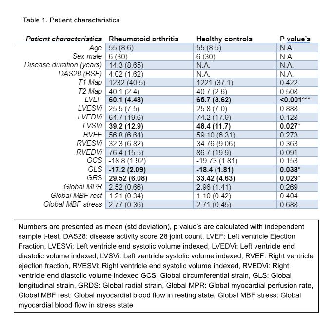
Results: Both LV global longitudinal strain (GLS), and LV global radial strain (GRS) were significantly reduced in RA patients as compared to healthy controls (p< 0.05 for both) (Table 1). RA patients also demonstrated a significant reduction in LVEF compared to healthy controls (60.1% in the RA patients compared to 65.7% in the healthy controls p< 0.001). T1 and T2 mapping were similar in both groups (1232 vs 1221 and 40.1 vs 40.7 respectively), No differences in myocardial perfusion imaging parameters were observed between the two groups. LGE identified fibrosis/scar was seen in 3 patients in the RA group, and not detected in the healthy volunteers.
Conclusion: Patients with RA, without (risk factors for) cardiovascular disease, have a reduced LV function as compared to healthy controls, as shown by a reduction in LVEF and LV strain. Other markers such as quantitative perfusion, T1 mapping, T2 mapping did not show any difference. Our results suggest screening for cardiac dysfunction might be indicated even in patients with no cardiac symptoms.
To cite this abstract in AMA style:
Dijkshoorn B, Mohamed S, Hansildaar R, Everaars H, Mooij G, Hopman L, Knaapen P, Robbers L, Nurmohamed M. Impaired Cardiac Function in Contemporary Rheumatoid Arthritis Patients: A Comparative Pilot Study with Cardiovascular Magnetic Resonance-derived Strain Analysis [abstract]. Arthritis Rheumatol. 2024; 76 (suppl 9). https://acrabstracts.org/abstract/impaired-cardiac-function-in-contemporary-rheumatoid-arthritis-patients-a-comparative-pilot-study-with-cardiovascular-magnetic-resonance-derived-strain-analysis/. Accessed .« Back to ACR Convergence 2024
ACR Meeting Abstracts - https://acrabstracts.org/abstract/impaired-cardiac-function-in-contemporary-rheumatoid-arthritis-patients-a-comparative-pilot-study-with-cardiovascular-magnetic-resonance-derived-strain-analysis/

