Session Information
Session Type: Poster Session C
Session Time: 9:00AM-11:00AM
Background/Purpose: Hypertrophic pachymeningitis (HP) is a rare inflammatory neurological disorder characterized by the thickened dura mater with extensive tissue fibrosis and immune-mediated inflammation. Notably, headaches and cranial neuropathies are frequently observed in immune-mediated HP, which is attributed to antineutrophil cytoplasmic antibody (ANCA)-associated vasculitis or IgG4-related disease as the common underlying diseases. However, it is still obscure how the volume and localization of HP lesions affect the development of the relevant neurological impairments. This study evaluated the quantification and localization of the thickened dura mater and analyzed their impact on clinical findings in immune-mediated HP.
Methods: We evaluated the volume of the contrast-enhanced dura mater on magnetic resonance imaging in 19 patients with HP, including 12 with ANCA-related, 4 with IgG4-related, and 3 with idiopathic HP, by the imaging feature quantification system. We enrolled 10 patients with multiple sclerosis (MS) as controls. In patients with HP, the impacts of HP volume on neurological symptoms and cerebrospinal fluid (CSF) laboratory markers were statistically analyzed. The receiver operating characteristic (ROC) curve analyses were also performed to detect the cut-off volume of the contrast-enhanced dura mater.
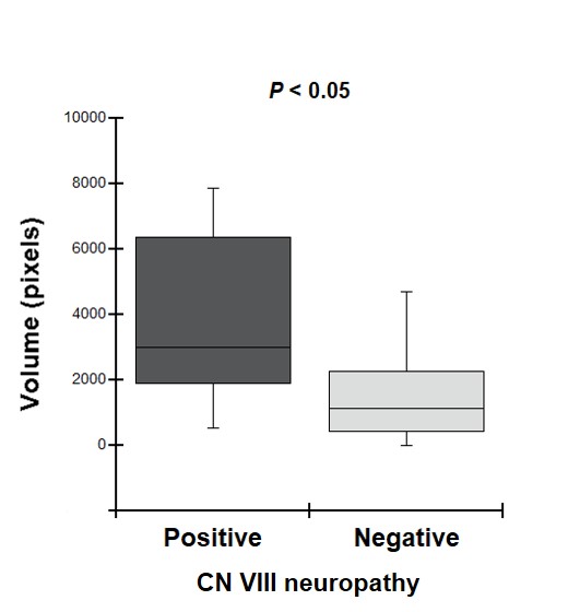
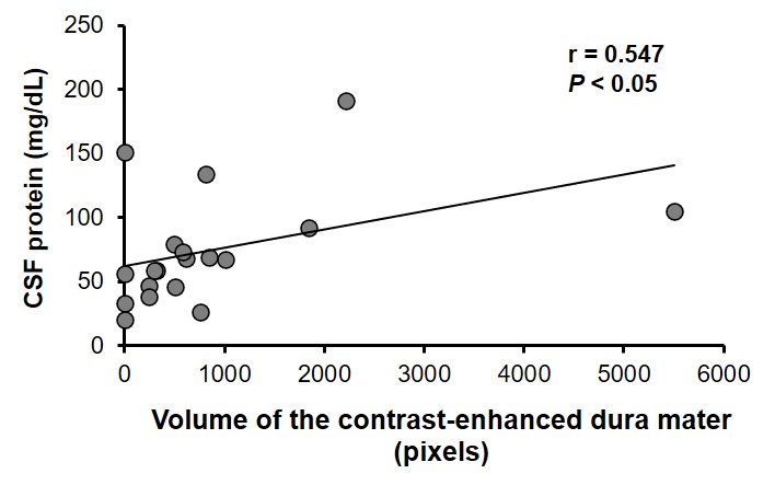
Results: Patients with HP demonstrated significantly higher volumes of the contrast-enhanced dura mater in the convexity, cranial fossa, and tentorium cerebelli than those with MS (P < 0.01). Of the 19 patients with HP, more than the cut-off volume in the convexity, cranial fossa, and tentorium cerebelli was observed in 16 (84%), 12 (63%), and 15 (79%) patients, respectively. Meanwhile, the volume of the contrast-enhanced dura mater was not significantly different among patients with ANCA-related, IgG4-related, and idiopathic HP. In patients with HP, those with cranial nerve (CN) VIII neuropathy had a significantly higher volume of the contrast-enhanced dura mater in the cranial fossa than those without CN VIII neuropathy (P < 0.05) (Fig. 1). The cut-off volume in the cranial fossa was more frequently exceeded in patients with CN VIII neuropathy than in those without CN VIII neuropathy. A positive correlation between the volume of the contrast-enhanced dura mater in the tentorium cerebelli and CSF protein levels was significantly observed in patients with HP (P < 0.05) (Fig. 2).
Conclusion: HP lesions in the cranial fossa may be robustly implicated in impairing CN VIII. The enlargement of HP lesions in the tentorium cerebelli can increase CSF protein levels. This study suggests that quantification of the thickened dura mater is useful for elucidating the relationship with the clinical findings in immune-mediated HP.
To cite this abstract in AMA style:
Shimojima Y, Ikeda J, Sekijima Y. Immune-mediated Hypertrophic Pachymeningitis: Focusing on the Localization and Volume of Thickened Dura Mater Lesion [abstract]. Arthritis Rheumatol. 2023; 75 (suppl 9). https://acrabstracts.org/abstract/immune-mediated-hypertrophic-pachymeningitis-focusing-on-the-localization-and-volume-of-thickened-dura-mater-lesion/. Accessed .« Back to ACR Convergence 2023
ACR Meeting Abstracts - https://acrabstracts.org/abstract/immune-mediated-hypertrophic-pachymeningitis-focusing-on-the-localization-and-volume-of-thickened-dura-mater-lesion/


