ACR Meeting Abstracts

ACR Meeting Abstracts

  • Meetings
    • ACR Convergence 2024
    • ACR Convergence 2023
    • 2023 ACR/ARP PRSYM
    • ACR Convergence 2022
    • ACR Convergence 2021
    • ACR Convergence 2020
    • 2020 ACR/ARP PRSYM
    • 2019 ACR/ARP Annual Meeting
    • 2018-2009 Meetings
    • Download Abstracts
  • Keyword Index
  • Advanced Search
  • Your Favorites
    • Favorites
    • Login
    • View and print all favorites
    • Clear all your favorites
  • ACR Meetings

Abstract Number: 1716

High-Throughput Semi-Automated Micro-CT Analysis Identifies the Cuboid Bone as a Sex-Dependent Biomarker of Inflammatory-Erosive Arthritis in TNF-Tg Mice

H. Mark Kenney1, Kiana Chen2, Lindsay Schnur3, Jeffrey Fox3, Ronald Wood3, Lianping Xing4, Christopher T Ritchlin5, Homaira Rahimi6, Edward Schwarz6 and Hani Awad3, 1University of Rochester School of Medicine and Dentistry, Henrietta, NY, 2University of Rochester School of Medicine and Dentistry, Rochester, NY, 3University of Rochester Medical Center, Rochester, NY, 4University of Rochester Medical Center, Webster, NY, 5University of Rochester Medical School, Allergy, Immunology & Rheumatology Division, Canandaigua, NY, 6University of Rochester, Rochester, NY

Meeting: ACR Convergence 2023

Keywords: Animal Model, Biomarkers, Computed tomography (CT), Erosions, Imaging

  • Tweet
  • Email
  • Print
Session Information

Date: Tuesday, November 14, 2023

Title: (1713–1733) RA – Animal Models Poster

Session Type: Poster Session C

Session Time: 9:00AM-11:00AM

Background/Purpose: Development of reliable disease activity biomarkers is critical for diagnostics, prognostics, and novel drug development. In the case of preclinical models of inflammatory-erosive arthritis, such as sexually dimorphic tumor necrosis factor transgenic (TNF-Tg) mice (1), disease severity is routinely quantified in the ankle through manual segmentation of the talus or small regions of adjacent bones primarily due to the ease in measurement (2,3). Herein, we sought to determine the particular hindpaw bones that represent reliable biomarkers of sex-dependent disease progression to guide future investigation and analysis.

Methods: Hindpaw micro-computed tomography (μCT) was performed on wild-type (n=4 male, n=4 female) and TNF-Tg (n=4 male, n=7 female) mice at monthly intervals from 2-5 (females) and 2-8-months (males) of age, where female TNF-Tg mice exhibit early mortality from cardiopulmonary disease at approximately 5-6-months (1). For image analysis, we utilized our recently developed high-throughput and semi-automated segmentation strategy in Amira software (v2020.2) (4). Synovial (H&E-OG) and osteoclast (TRAP) areas of ankle joints were quantified using Visiopharm (v2021.07).

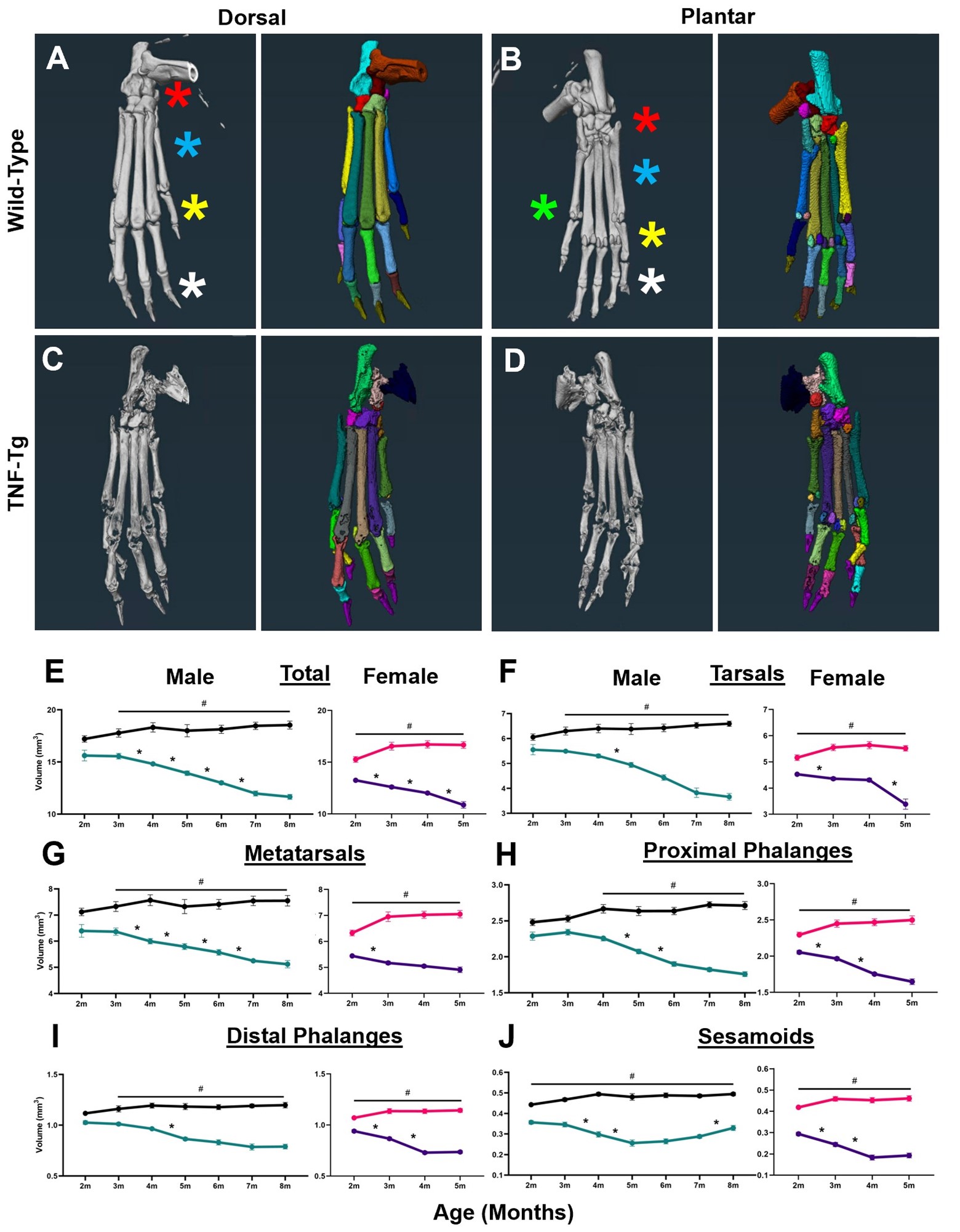
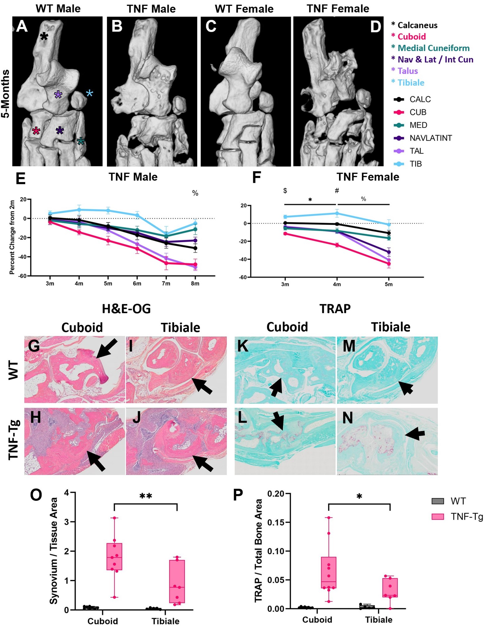
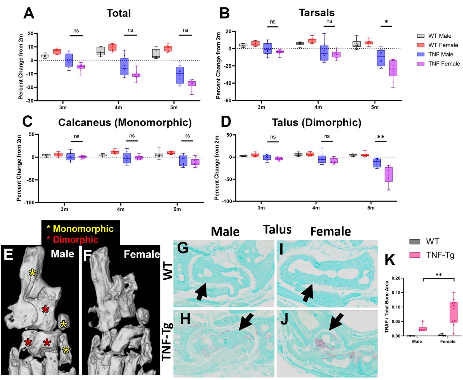
Results: First, we demonstrated our analysis method had comparable automated segmentation accuracy in wild-type and TNF-Tg hindpaws before correction (79.2±8.9% vs 80.1±5.1%, p=0.52), determined through analysis of ~9000 individual bones by a single user, with representative μCT (left) and bone segmentation (right) provided (Fig. 1A-D). Compared to other bone compartments, the tarsal region demonstrated a sudden, specific, and significant bone volume reduction in female TNF-Tg mice by 5-months (Fig. 1E-J). This sexual dimorphism was associated with unique bone-specific changes across time, as the cuboid at 4-months of age showed significantly reduced bone volumes compared to all other tarsals. In contrast, TNF-Tg male mice exhibited no difference between individual bone volumes at this timepoint (Fig. 2A-F). Compared to bones with limited erosions (i.e., tibiale), the cuboid showed a corresponding increased synovial and TRAP area (Fig. 2G-P). At 5-months of age, additional bones localized to the antero-lateral region of the ankle were also responsible for the dramatic erosions in the tarsal region of females (Fig. 3A-F, red stars), coinciding with increased TRAP+ osteoclasts in female vs male TNF-Tg mice (Fig. 3G-K).

Conclusion: Taken together, here we demonstrated that sexual dimorphism of arthritis in TNF-Tg mice is bone-specific, where the cuboid serves as a reliable biomarker of erosive arthritis with the greatest sensitivity to early and consistent bone loss related to enhanced osteoclast numbers. Ongoing work will further investigate the cellular mechanisms of these bone-specific erosions in mice and evaluate the translational potential of these biomarkers in arthritis patients through our segmentation model. 1. Bell et al. Arthritis Rheumatol. 71(9):1512-1523. 2019. 2. Proulx et al. Arthritis Rheumatol. 56(12):4024-4037. 2007. 3. Cambre et al. Nat Commun. 9(1):4613. 2018. 4. Kenney et al. Bone Rep. 16(101167). 2022.

Supporting image 1

Figure 1. Semi-automated segmentation of TNF-Tg hindpaws reveals unique temporal patterns of erosions by bone compartment in female mice. To evaluate bone-specific erosions in the TNF-Tg mouse model of inflammatory-erosive arthritis, we utilized our recently published high-throughput semi-automated hindpaw segmentation protocol (4). Representative images of the dorsal and plantar surfaces of hindpaw micro-CT images (left) and the segmentation of each bone indicated by unique colors (right) are provided for wild-type (A, B) and TNF-Tg (C, D) male mice at 8-months of age. Without user intervention, the semi-automated protocol produced accurate segmentations of approximately 80% of bones in wild-type datasets (error rate ~20%), which remarkably remains consistent, and potentially improved due to reduced variance, for TNF-Tg mice with bone erosions. Patterns of bone loss in hindpaw compartments (shown in A, B: tarsals, red star; metatarsals, blue; proximal phalanges, yellow; distal phalanges, white; and sesamoids, green) were analyzed for temporal and genotype effects. Although both TNF-Tg males and females exhibited consistent bone loss across time (E, *p<0.05), TNF-Tg females showed unique time-dependent erosions by particular compartments. For TNF-Tg females, there was a dramatic decrease in the bone volume of the tarsals between 4-5-months of age (F) that was preceded by early erosions starting at 2-months of age in the bones associated with the phalanges that was not sustained past 4-months (G-J). In contrast, males showed relatively slow progression of erosions with limited statistical change for particular compartments at monthly intervals, except for the metatarsals (F-J). Also note the “U”-shaped progression of bone volumes in the TNF-Tg male sesamoids, likely representing reactive bone remodeling following an initial period of erosions (J). Compared to wild-type, both TNF-Tg male and female mice showed significantly reduced bone volumes in all compartments starting at 3-months and 2-months of age, respectively (#p<0.05). Statistics: 2-way ANOVA (males) and mixed-effects analysis (females) with Tukey’s multiple comparisons (E-J).

Supporting image 2

Figure 2. The cuboid is an early biomarker of erosive arthritis in TNF-Tg female mice. To further elucidate the dynamic sex-dependent, bone-specific, and temporal changes in the tarsals of TNF-Tg mice, we directly compared erosive activity between each tarsal bone to determine their biomarker potential during progression of inflammatory arthritis. High-magnification images of the tarsal compartment at 5-months of age, with the individual bones indicated by stars (calcaneus, black; cuboid, pink; medial cuneiform, green; fused navicular, lateral and intermediate cuneiform [a known C57BL/6 murine variant (4)], dark purple; talus, light purple; and tibiale, blue), are shown for each group (A-D). In order to compare the individual bones, percent change in bone volume from their baseline at 2-months was calculated to control for the differences in bone size. TNF-Tg males exhibited no significant difference in erosions between individual bones in the tarsal region, except for the tibiale, which showed a resistance to erosions compared to all other bones, except the medial cuneiform (% p<0.05) (E). In contrast, TNF-Tg females exhibited early erosive activity localized specifically to the cuboid bone with significantly reduced bone volumes compared to the other bones in the tarsal region by 3- and 4-months of age ($ cuboid vs all bones except medial cuneiform, # cuboid vs all bones, p<0.05). In addition, significant decreases in bone volume over time from 3- to 4-months of age were noted only in the cuboid and fused navicular, lateral cuneiform and intermediate cuneiform (* p<0.05). All bones, except for the tibiale, showed significantly reduced bone volumes from 4- to 5-months of age (% p<0.05), explaining the time-dependent erosions of the tarsal compartment during this timeframe, noted in Fig. 1 (F). To understand the cellular mechanisms mediating resistance (i.e., tibiale) and susceptibility (i.e., cuboid) to erosions, we histologically assessed and quantified the amount of synovium (H&E-OG) and osteoclasts (TRAP) surrounding these particular bones (arrows). Compared to the tibiale, the cuboid exhibited both increased synovial infiltrate (G-J, purple) and TRAP+ osteoclasts (K-N, red), likely involved in mediating the bone-specific erosions (G-P). Statistics: 2-way ANOVA (E, O, P) and mixed effects analysis (F) with Tukey’s multiple comparisons.

Supporting image 3

Figure 3. Anterolateral tarsal bones show severe female-specific erosions in TNF-Tg mice. Sex-dependent differences in bone erosions within the tarsal compartment were evaluated using percent change in bone volumes to normalize for animal size between groups. While total bone volume showed no significant changes between TNF-Tg males and females (A), the tarsal compartment exhibited reduced bone volumes in females compared to males at 5-months of age (B). Interestingly, for bones such as the calcaneus (C), there were no changes noted between the sexes. The significant reduction in tarsal bone volume in females was instead driven by sex-specific erosive activity in bones such as the talus (D). The particular bones associated with erosion of the tarsals in females are notably localized to the anterolateral region of the ankle (red stars), while those without sexual dimorphism are oriented posteromedially (yellow stars). Representative micro-CT images of male (E) and female (F) TNF-Tg mice at 5-months of age exhibit the lateralization of the sexually dimorphic effects (reproduced from Fig. 2 for convenience). Corresponding with these bone-specific changes, histologic evaluation revealed significantly increased TRAP+ osteoclasts (red) around the talus in TNF-Tg females vs males at 5-months of age (G-K). Statistics: Mixed effects analysis (A-D) and 2-way ANOVA (K) with Tukey’s multiple comparisons; *p<0.05, **p<0.01.


Disclosures: H. Kenney: None; K. Chen: None; L. Schnur: None; J. Fox: None; R. Wood: None; L. Xing: None; C. Ritchlin: AbbVie, 2, 5, 6, Amgen, 2, BMS, 2, Eli Lilly, 2, Gilead, 2, Janssen, 2, Novartis, 2, Pfizer, 2, 5, 6, UCB, 2, 6; H. Rahimi: None; E. Schwarz: None; H. Awad: None.

To cite this abstract in AMA style:

Kenney H, Chen K, Schnur L, Fox J, Wood R, Xing L, Ritchlin C, Rahimi H, Schwarz E, Awad H. High-Throughput Semi-Automated Micro-CT Analysis Identifies the Cuboid Bone as a Sex-Dependent Biomarker of Inflammatory-Erosive Arthritis in TNF-Tg Mice [abstract]. Arthritis Rheumatol. 2023; 75 (suppl 9). https://acrabstracts.org/abstract/high-throughput-semi-automated-micro-ct-analysis-identifies-the-cuboid-bone-as-a-sex-dependent-biomarker-of-inflammatory-erosive-arthritis-in-tnf-tg-mice/. Accessed .
  • Tweet
  • Email
  • Print

« Back to ACR Convergence 2023

ACR Meeting Abstracts - https://acrabstracts.org/abstract/high-throughput-semi-automated-micro-ct-analysis-identifies-the-cuboid-bone-as-a-sex-dependent-biomarker-of-inflammatory-erosive-arthritis-in-tnf-tg-mice/

Advanced Search

Your Favorites

You can save and print a list of your favorite abstracts during your browser session by clicking the “Favorite” button at the bottom of any abstract. View your favorites »

All abstracts accepted to ACR Convergence are under media embargo once the ACR has notified presenters of their abstract’s acceptance. They may be presented at other meetings or published as manuscripts after this time but should not be discussed in non-scholarly venues or outlets. The following embargo policies are strictly enforced by the ACR.

Accepted abstracts are made available to the public online in advance of the meeting and are published in a special online supplement of our scientific journal, Arthritis & Rheumatology. Information contained in those abstracts may not be released until the abstracts appear online. In an exception to the media embargo, academic institutions, private organizations, and companies with products whose value may be influenced by information contained in an abstract may issue a press release to coincide with the availability of an ACR abstract on the ACR website. However, the ACR continues to require that information that goes beyond that contained in the abstract (e.g., discussion of the abstract done as part of editorial news coverage) is under media embargo until 10:00 AM ET on November 14, 2024. Journalists with access to embargoed information cannot release articles or editorial news coverage before this time. Editorial news coverage is considered original articles/videos developed by employed journalists to report facts, commentary, and subject matter expert quotes in a narrative form using a variety of sources (e.g., research, announcements, press releases, events, etc.).

Violation of this policy may result in the abstract being withdrawn from the meeting and other measures deemed appropriate. Authors are responsible for notifying colleagues, institutions, communications firms, and all other stakeholders related to the development or promotion of the abstract about this policy. If you have questions about the ACR abstract embargo policy, please contact ACR abstracts staff at [email protected].

Wiley

  • Online Journal
  • Privacy Policy
  • Permissions Policies
  • Cookie Preferences

© Copyright 2025 American College of Rheumatology