
Session Information
Date: Monday, November 18, 2024
Title: Muscle Biology, Myositis & Myopathies – Basic & Clinical Science Poster III
Session Type: Poster Session C
Session Time: 10:30AM-12:30PM
Background/Purpose: Recently, the role of imaging tests such as magnetic resonance imaging and ultrasound (US) in the diagnosis and classification of myositis has become increasingly important. However, the classification of myositis, particularly distinguishing inclusion body myositis (IBM) from non-IBM cases by US, remains challenging. Traditional methods rely on qualitative expert analysis, but quantitative techniques such as echo intensity (EI) and texture analysis have emerged as potential tools for improving diagnostic accuracy. This study aims to evaluate the efficacy of texture analysis and EI in myositis classification and IBM/non-IBM differentiation and to compare these methods with expert qualitative analysis.
Methods: From 2014 to 2023, we cross-sectionally evaluated patients with myositis who were admitted to our network institutions with a new diagnosis or significant flare of myositis. We collected clinical information and laboratory test results, as well as US images. Quantitative measures included EI, histogram parameters (skewness and kurtosis), and texture parameters such as contrast, entropy, angular second moment, correlation, and inverse difference moment. The texture parameters were measured at different angles (0° or 90°) and interpixel distances (1, 3, and 5 pixels), resulting in six measurements in each muscle. These analyses were applied to the biceps brachii (BB) and vastus medialis (VM) muscles using the same US machine/settings. ImageJ, an image processing software, was used for the measurement of EI and texture parameters. Logistic regression models were developed for myositis vs. control and IBM vs. non-IBM classifications. Additionally, two experts in muscle ultrasound independently evaluated the images for qualitative analysis. Cases of disagreement were resolved through consensus or consultation with a senior US specialist.
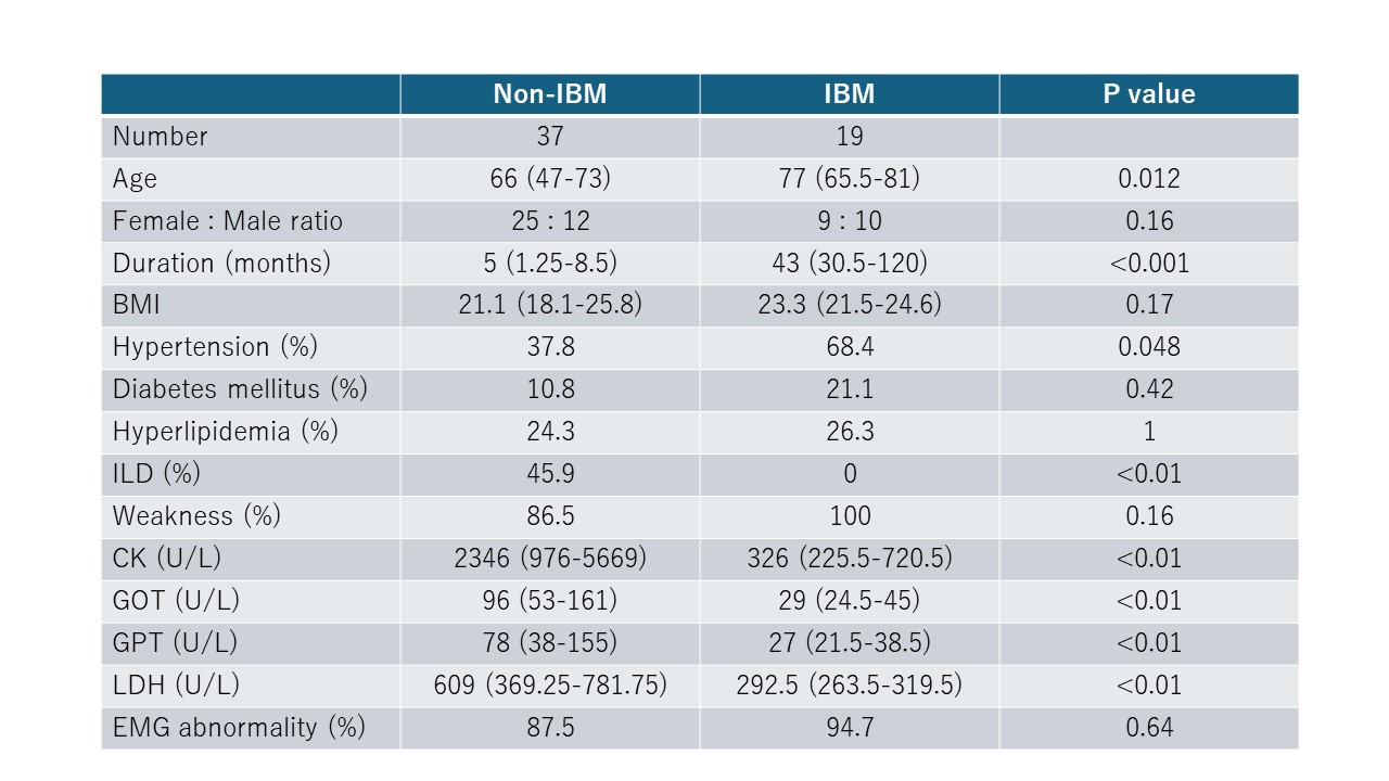
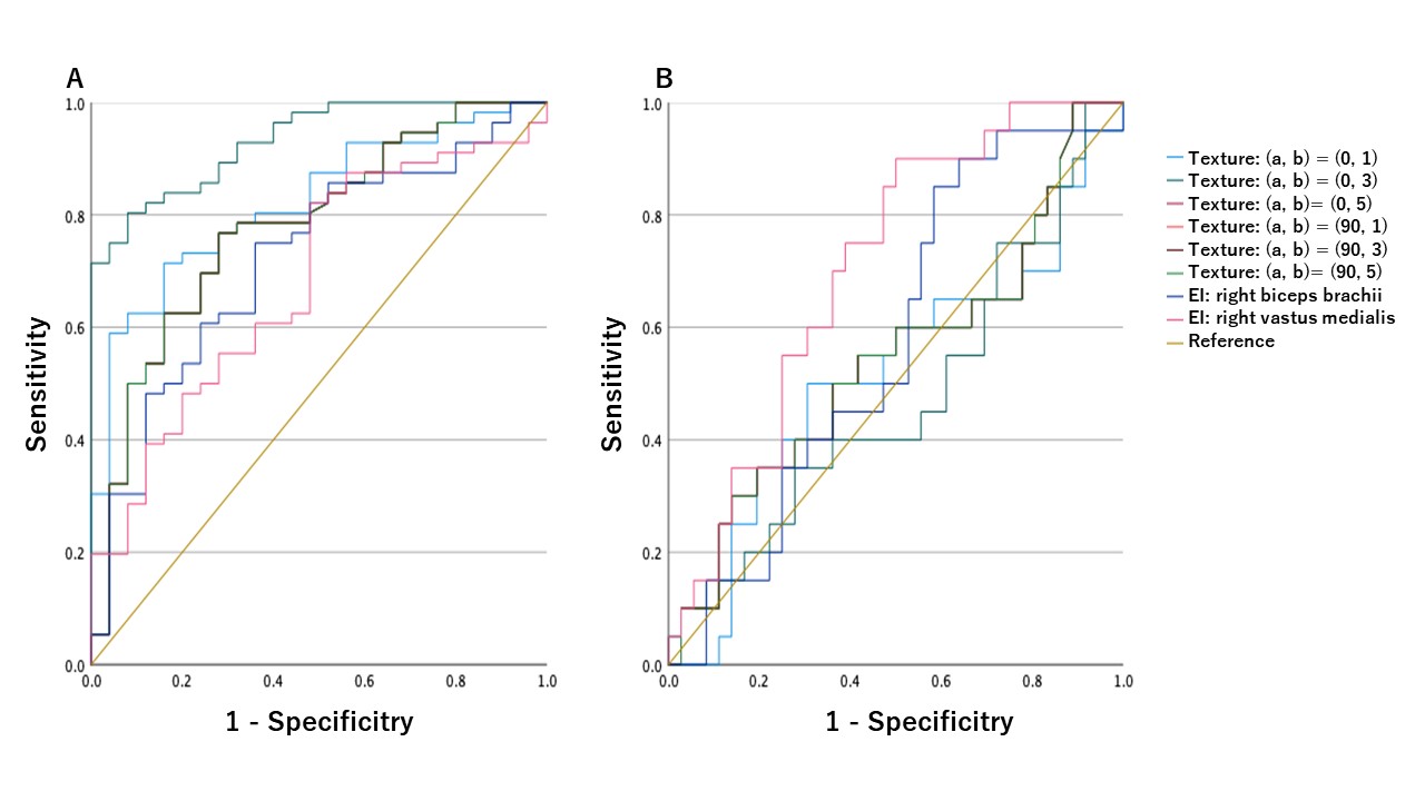
Results: A total of 56 patients with myositis (37 non-IBM, 19 IBM) and 25 age/sex-matched inflammatory controls (patients with rheumatoid arthritis) underwent ultrasound evaluation. Baseline characteristics showed that IBM patients were older, had less F:M ratio, and had less CK and other muscle enzyme levels. In the differentiation of myositis from controls, texture analysis significantly outperformed EI alone, with a sensitivity of 80.4% and specificity of 92%. For IBM vs. non-IBM classification, EI provided better performance (sensitivity 70%, specificity 63.9%) compared to texture analysis. The expert qualitative analysis achieved a sensitivity of 82.5% and specificity of 83.3% for myositis vs. control, and 94.7% and 79% for IBM vs. non-IBM, respectively.
Conclusion: Texture analysis enhances the differentiation of myositis from controls, outperforming EI. However, for IBM vs. non-IBM classification, EI remains superior. Expert qualitative analysis shows comparable to better performance compared to quantitative methods, highlighting the value of expert experience in muscle ultrasound. These findings suggest that the addition of texture analysis to EI could optimize myositis classification, but expert qualitative assessment still holds significant value in clinical practice.
To cite this abstract in AMA style:
Yoshida T, Yamazaki H, Takamatsu N, Taniguchi Y, Nozaki T, inoue m, Kumon Y, matsumoto y, Kanazawa Y, Harada M, Nishino I, Albayda J, Izumi Y. Comparative Diagnostic Efficacy of Quantitative Measurement of Echo Intensity and Texture versus Qualitative Analysis of Ultrasound Images in Myositis [abstract]. Arthritis Rheumatol. 2024; 76 (suppl 9). https://acrabstracts.org/abstract/comparative-diagnostic-efficacy-of-quantitative-measurement-of-echo-intensity-and-texture-versus-qualitative-analysis-of-ultrasound-images-in-myositis/. Accessed .ACR Meeting Abstracts - https://acrabstracts.org/abstract/comparative-diagnostic-efficacy-of-quantitative-measurement-of-echo-intensity-and-texture-versus-qualitative-analysis-of-ultrasound-images-in-myositis/