ACR Meeting Abstracts

ACR Meeting Abstracts

  • Meetings
    • ACR Convergence 2024
    • ACR Convergence 2023
    • 2023 ACR/ARP PRSYM
    • ACR Convergence 2022
    • ACR Convergence 2021
    • ACR Convergence 2020
    • 2020 ACR/ARP PRSYM
    • 2019 ACR/ARP Annual Meeting
    • 2018-2009 Meetings
    • Download Abstracts
  • Keyword Index
  • Advanced Search
  • Your Favorites
    • Favorites
    • Login
    • View and print all favorites
    • Clear all your favorites
  • ACR Meetings

Abstract Number: 0479

A Novel Semi-automated Classifier of Radiographic Hip Osteoarthritis on DXA Scans Is Strongly Predictive of Pain, Clinical Diagnosis and Joint Replacement: Findings from 40,000 Participants in UK Biobank

Benjamin Faber1, Raja Ebsim2, Fiona Saunders3, Monika Frysz1, Claudia Lindner2, Jennifer Gregory3, Richard Aspden3, Nicholas Harvey4, George Davey Smith1, Timothy Cootes2 and Jon Tobias1, 1University of Bristol, Bristol, United Kingdom, 2University of Manchester, Manchester, United Kingdom, 3University of Aberdeen, Aberdeen, United Kingdom, 4University of Southampton, Southampton, United Kingdom

Meeting: ACR Convergence 2021

Keywords: Epidemiology, Osteoarthritis, pain, radiography, Total joint replacement

  • Tweet
  • Email
  • Print
Session Information

Date: Saturday, November 6, 2021

Title: Abstracts: Osteoarthritis – Clinical (0478–0483)

Session Type: Abstract Session

Session Time: 2:15PM-2:30PM

Background/Purpose: Radiographic hip osteoarthritis (rHOA) is traditionally defined on hip or pelvic radiographs, using subjective methods such as Kellgren-Lawrence scoring. Associations between subjective rHOA measures and clinical outcomes such as hip pain are inconsistent. Applying digital analytical tools to high-resolution dual-energy X-ray absorptiometry (DXA) scans, we aimed to (1) develop a novel semi-automated classifier for rHOA, (2) apply this to available hip DXAs from UK Biobank (UKB), and (3) evaluate face validity of the classifier based on prevalence data and relationships with hip pain, hospital diagnosed OA, and risk of total hip replacement (THR).

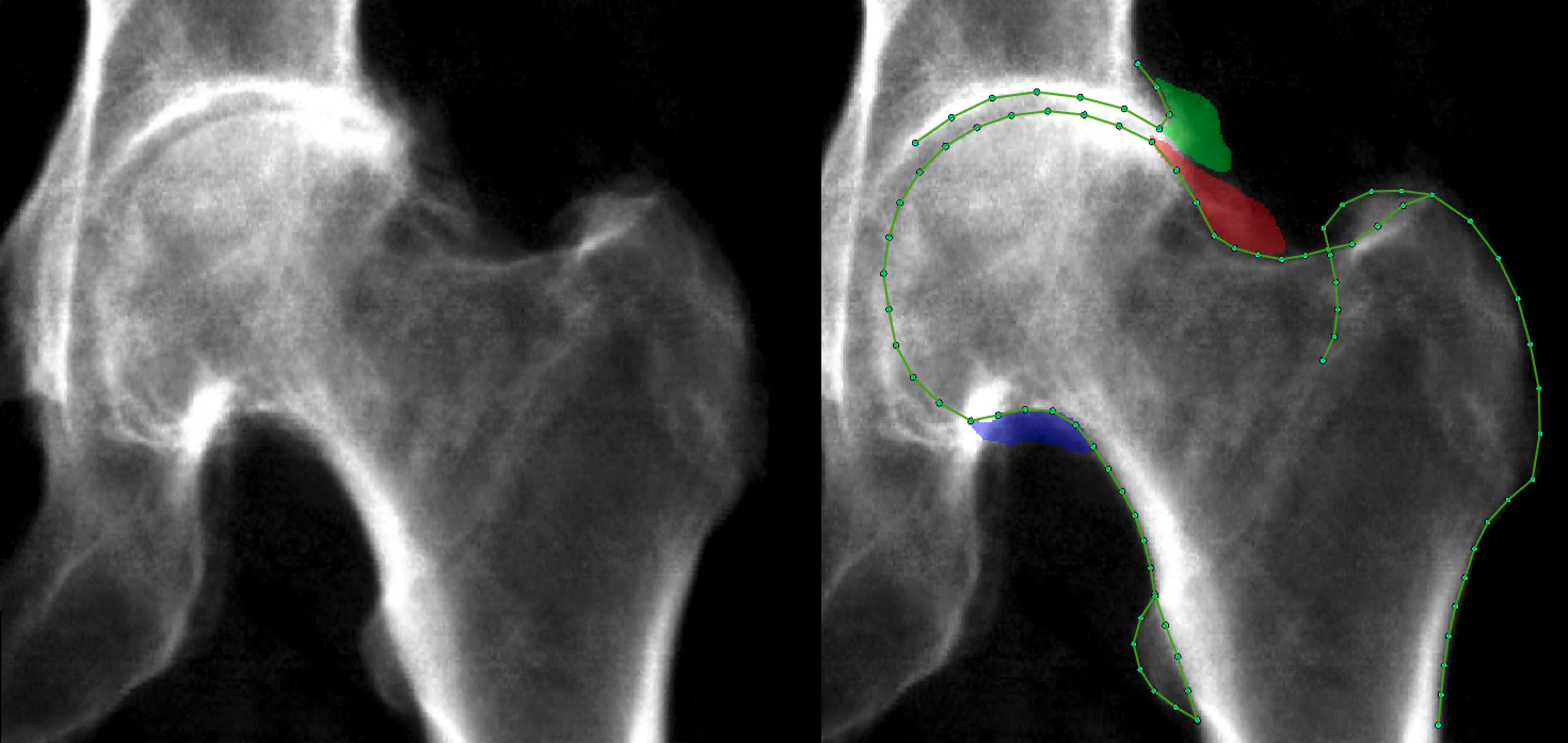
Methods: Osteophyte area was manually measured (in mm2) at the lateral acetabulum, superior-lateral and inferior-medial femoral head on left hip DXAs obtained from individuals in UKB (Figure 1). Osteophyte area thresholds were defined and subsequently applied to categorise each osteophyte into grades 0-3. Minimum joint space width (mJSW) was automatically measured (in mm) over the superior joint space using outline points placed by a machine learning based approach. Thresholds of mJSW were used to categorise individuals into joint space narrowing (JSN) grades 0-3. Osteophyte and JSN grades were summated for each individual to give a rHOA score of 0-12. Hips were then categorised into rHOA grades 0-4 based on rHOA score. Logistic regression was used to examine associations between rHOA grade and hip pain derived from a questionnaire, and hospital diagnosed OA and THR from electronic health record national linkage (HES). Our adjusted model included age, sex, height and weight as covariates.

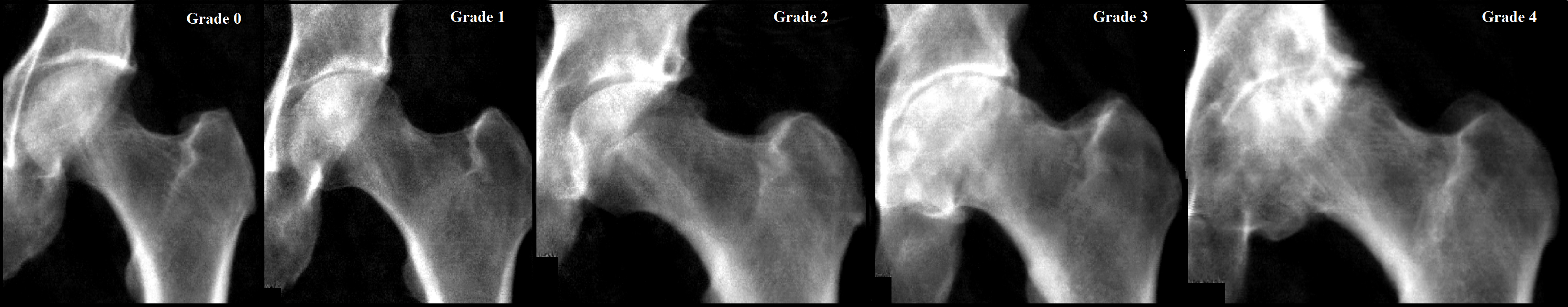
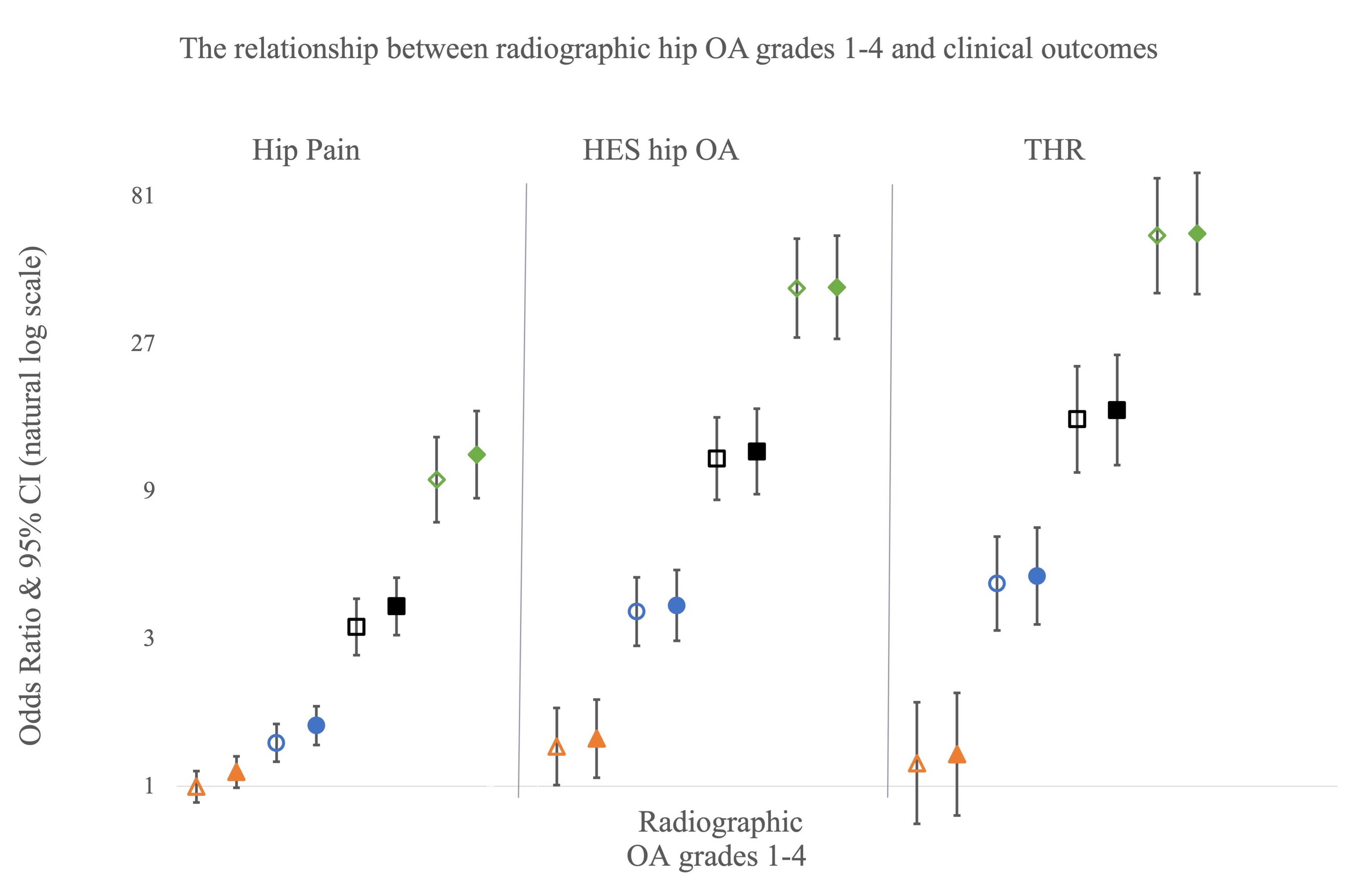
Results: 40,340 individuals were included in the study [mean age 63.7 (range 44-82), 19294/21046 male/female]. 32758 (81.2%) had rHOA grade 0, 4565 (11.3%) grade 1, 2317 (5.7%) grade 2, 543 (1.3%) grade 3, 157 (0.4%) grade 4 (Figure 2), with all features of rHOA being more common in males than females. rHOA grades ≥2 were associated with all three clinical outcomes in both unadjusted and adjusted models, a clear dose-response relationship was seen with each increase in grade showing a large rise in odds ratio (Figure 3).

Conclusion: Having developed a semi-automated classifier for rHOA on hip DXA scans, we successfully applied this to over 40,000 individuals from UKB, yielding expected findings for OA prevalence including higher rates in males compared with females. The validity of our classifier was further supported by the strong and progressive relationships observed between rHOA and hip pain, and risks of hospital diagnosed hip OA and THR. We conclude that hip DXA scans provide a promising means of objectively defining rHOA, with potential application in the clinic and as a research tool.

Figure 1. The left image shows a DXA scan from UK Biobank. The right image shows the same DXA scan with osteophytes and outline points marked.

Figure 2. Example DXAs of each rHOA grade

Figure 3. The associations between rHOA grades (1_4) and clinical outcomes. Odds ratios are plotted with 95% confidence intervals either side. Triangles = grade 1, circles = grade 2, squares = grade 3, and diamonds = grade 4. Unadjusted results = hollow shapes, and adjusted results = filled shapes. The Y-axis scale is natural log based.


Disclosures: B. Faber, None; R. Ebsim, None; F. Saunders, None; M. Frysz, None; C. Lindner, None; J. Gregory, None; R. Aspden, None; N. Harvey, UCB, 2, 6, Kyowa Kirin, 2, 6, Internis Pharma, 2, 6; G. Davey Smith, None; T. Cootes, None; J. Tobias, None.

To cite this abstract in AMA style:

Faber B, Ebsim R, Saunders F, Frysz M, Lindner C, Gregory J, Aspden R, Harvey N, Davey Smith G, Cootes T, Tobias J. A Novel Semi-automated Classifier of Radiographic Hip Osteoarthritis on DXA Scans Is Strongly Predictive of Pain, Clinical Diagnosis and Joint Replacement: Findings from 40,000 Participants in UK Biobank [abstract]. Arthritis Rheumatol. 2021; 73 (suppl 9). https://acrabstracts.org/abstract/a-novel-semi-automated-classifier-of-radiographic-hip-osteoarthritis-on-dxa-scans-is-strongly-predictive-of-pain-clinical-diagnosis-and-joint-replacement-findings-from-40000-participants-in-uk-biob/. Accessed .
  • Tweet
  • Email
  • Print

« Back to ACR Convergence 2021

ACR Meeting Abstracts - https://acrabstracts.org/abstract/a-novel-semi-automated-classifier-of-radiographic-hip-osteoarthritis-on-dxa-scans-is-strongly-predictive-of-pain-clinical-diagnosis-and-joint-replacement-findings-from-40000-participants-in-uk-biob/

Advanced Search

Your Favorites

You can save and print a list of your favorite abstracts during your browser session by clicking the “Favorite” button at the bottom of any abstract. View your favorites »

All abstracts accepted to ACR Convergence are under media embargo once the ACR has notified presenters of their abstract’s acceptance. They may be presented at other meetings or published as manuscripts after this time but should not be discussed in non-scholarly venues or outlets. The following embargo policies are strictly enforced by the ACR.

Accepted abstracts are made available to the public online in advance of the meeting and are published in a special online supplement of our scientific journal, Arthritis & Rheumatology. Information contained in those abstracts may not be released until the abstracts appear online. In an exception to the media embargo, academic institutions, private organizations, and companies with products whose value may be influenced by information contained in an abstract may issue a press release to coincide with the availability of an ACR abstract on the ACR website. However, the ACR continues to require that information that goes beyond that contained in the abstract (e.g., discussion of the abstract done as part of editorial news coverage) is under media embargo until 10:00 AM ET on November 14, 2024. Journalists with access to embargoed information cannot release articles or editorial news coverage before this time. Editorial news coverage is considered original articles/videos developed by employed journalists to report facts, commentary, and subject matter expert quotes in a narrative form using a variety of sources (e.g., research, announcements, press releases, events, etc.).

Violation of this policy may result in the abstract being withdrawn from the meeting and other measures deemed appropriate. Authors are responsible for notifying colleagues, institutions, communications firms, and all other stakeholders related to the development or promotion of the abstract about this policy. If you have questions about the ACR abstract embargo policy, please contact ACR abstracts staff at [email protected].

Wiley

  • Online Journal
  • Privacy Policy
  • Permissions Policies
  • Cookie Preferences

© Copyright 2025 American College of Rheumatology