Session Information
Session Type: Abstract Session
Session Time: 12:30PM-12:45PM
Background/Purpose: To characterize muscle involvement in idiopathic inflammatory myopathies (IIMs) using detailed anatomical segmentation and volumetric 18F-FDG PET/CT analysis, and to evaluate the prognostic value of global metabolic burden compared to SUVmax.
Methods: Patients with IIMs undergoing baseline 18F-FDG PET/CT were retrospectively analyzed. Clinical and laboratory data (CK, AST, LDH, CRP, ESR) were collected. Severe or refractory disease was defined clinically. Each muscle group was individually assessed, then categorized into five regions (head/neck, thorax, abdomen, upper and lower extremities) based on anatomical curricula. PET parameters included SUVmax (peak activity), metabolic lesion volume (MLV), and total lesion glycolysis (TLG; SUVmean × MLV). Log-transformation normalized data. Pearson correlations and nonparametric tests assessed associations. ROC analysis after logistic regression evaluated prognostic performance.
Results: Twenty-one patients [median age 62 (range 22–77); 76.2% female] were included. Diagnoses were dermatomyositis (71.4%), polymyositis (23.8%), and antisynthetase syndrome (4.8%). Median follow-up was 11.5 months. Detailed anatomical segmentation (Table 1) revealed that thoracic muscles, particularly autochthonous thoracic and thoracic wall muscles, had the highest MLV and TLG values (median TLG: 115.99 thorax vs. 127.5 lower extremities). Lower extremities also showed significant burden, whereas abdominal and head/neck muscles were less involved (Figure 1). At follow-up, 33.3% (7/21) developed severe disease and 42.9% (9/21) were refractory. SUVmax did not correlate significantly with baseline laboratory markers or outcomes. In contrast, TLG demonstrated strong correlations with AST (r=0.657, p=0.003), LDH (r=0.663, p=0.003), CRP (r=0.598, p=0.009), and ESR (r=0.549, p=0.018). ROC analysis showed that TLG (AUC 0.821) and MLV (AUC 0.803) outperformed SUVmax (AUC 0.652) in predicting severe/refractory disease (Figure 2).
Conclusion: This study provides the first detailed volumetric map of muscle inflammation in IIMs using 18F-FDG PET/CT.Thoracic and paraspinal muscles were major sites of involvement, challenging the classical focus on proximal muscles. Volumetric PET parameters, particularly TLG and MLV, more effectively captured the global inflammatory load and correlated with severe or refractory disease. These findings suggest that comprehensive volumetric PET/CT analysis may enhance diagnostic precision and risk stratification in IIMs.
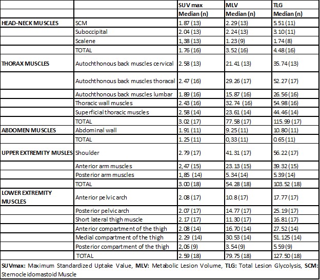 Table 1. Distribution of Muscle Involvement by Muscle Groups in Idiopathic Inflammatory Myopathy Patients as Assessed by 18F-FDG PET/CT (SUVmax, MLV, and TLG).
.jpg) Figure 1.Graphical Distribution of Muscle Group Involvement in IIM Patients Based on MLV and TLG Measurements.
Figure 1.Graphical Distribution of Muscle Group Involvement in IIM Patients Based on MLV and TLG Measurements.
.jpg) Figure 2. ROC Curves Comparing the Discriminative Ability of SUVmax, MLV, and TLG for Predicting Severe or Refractory Disease.
Figure 2. ROC Curves Comparing the Discriminative Ability of SUVmax, MLV, and TLG for Predicting Severe or Refractory Disease.
To cite this abstract in AMA style:
Basibuyuk F, Bahadir M, Kenar Artin G, Kiray A, Bekis R, Sari İ. Quantitative 18F-FDG PET/CT analysis reveals new patterns of muscle involvement and prognostic indicators in idiopathic inflammatory myopathies: beyond proximal muscle weakness [abstract]. Arthritis Rheumatol. 2025; 77 (suppl 9). https://acrabstracts.org/abstract/quantitative-18f-fdg-pet-ct-analysis-reveals-new-patterns-of-muscle-involvement-and-prognostic-indicators-in-idiopathic-inflammatory-myopathies-beyond-proximal-muscle-weakness/. Accessed .« Back to ACR Convergence 2025
ACR Meeting Abstracts - https://acrabstracts.org/abstract/quantitative-18f-fdg-pet-ct-analysis-reveals-new-patterns-of-muscle-involvement-and-prognostic-indicators-in-idiopathic-inflammatory-myopathies-beyond-proximal-muscle-weakness/