Session Information
Date: Monday, October 27, 2025
Title: (0955–0977) Systemic Sclerosis & Related Disorders – Basic Science Poster I
Session Type: Poster Session B
Session Time: 10:30AM-12:30PM
Background/Purpose: Systemic Sclerosis (SSc) is a rare autoimmune connective tissue disease and presents a significant medical challenge. It is characterized by fibrotic tissue remodeling, along with microvascular alterations and autoimmunity with the formation of specific autoantibodies. CD19- and BCMA-targeted B cell depletion therapies such as CAR-T cells or bispecific T cell engaging antibodies demonstrated profound efficacy for the treatment of SSc patients, including those who were previously unresponsive to multiple therapies. The mechanisms underlying B cell activation in target tissues of SSc patients are largely unknown. Our study focuses on characterizing immune cell infiltration and the cellular microenvironment in SSc skin, with a particular interest in the formation of tertiary lymphoid structures.
Methods: Skin punch biopsies from SSc patients were formalin-fixed, paraffin-embedded, and sectioned. Sections of 118 patients with SSc were screened by hematoxylin and eosin (H&E) histological staining for the presence of leukocyte aggregates or tertiary lymphoid structures. Ten representative samples containing tertiary lymphoid structures, six samples negative for lymphoid infiltrates and five samples derived from normal healthy skin were stained using metal isotope labeled antibodies targeting a panel of proteins of interest including markers of immune and stromal cell populations. The staining protocol was established using human tonsil sections. Imaging Mass Cytometry (IMC) was performed on the Hyperion platform (Standard BioTools) to collect data on the spatial distribution of protein targets and co-localization of cell populations within the skin tissue.
Results: Tertiary lymphoid structures were detected in 14 out of 118 skin samples (12%) (Figure 1). Large, diffuse lymphoid infiltrates were recognized in 19 Sections (16%) while 13 sections (11%) were negative for lymphoid infiltrates.We were able to identify multiple stromal and immune cell clusters using our IMC panel with specific phenotypes of fibroblasts and endothelial cells in tertiary lymphoid structures. Immune cells were found to infiltrate defined stromal cell niches in the skin. Neighborhood analyses demonstrated context-specific interactions of leukocytes with stromal cells in these tertiary lymphoid structures. We will further analyze the demonstrated specific cellular proximity and co-localization, with a focus on mechanisms potentially involved in immune activation and inflammation in SSc.
Conclusion: Tertiary lymphoid structures and large, diffuse lymphoid infiltrates were detected in nearly one third of the SSc samples that were screened (28%), while only 11% of samples were negative for infiltrates. Using a state-of-the-art technique such as IMC, we provide first data on the cellular composition, the topographical organization and the cellular interactions with tertiary lymphoid structures in SSc. These data may enable targeted inhibition of the formation of these structures and thereby interference with tissue resident immune responses in SSc.
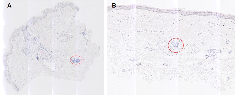 Figure 1: H&E stainings of tertiary lymphoid structures and large, diffuse lymphoid infiltrates in SSc skin sections. Exemplary images of tertiary lymphoid structures (A) and large, diffuse lymphoid infiltrates (B) in H&E stainings of SSc skin sections.
To cite this abstract in AMA style:
Bleck D, Drechsel K, Filla T, Li Y, Györfi A, Matei A, Distler J. Spatial Proteomics Analysis of the organization of tertiary lymphoid structures in Systemic Sclerosis Skin [abstract]. Arthritis Rheumatol. 2025; 77 (suppl 9). https://acrabstracts.org/abstract/spatial-proteomics-analysis-of-the-organization-of-tertiary-lymphoid-structures-in-systemic-sclerosis-skin/. Accessed .« Back to ACR Convergence 2025
ACR Meeting Abstracts - https://acrabstracts.org/abstract/spatial-proteomics-analysis-of-the-organization-of-tertiary-lymphoid-structures-in-systemic-sclerosis-skin/