Session Information
Date: Monday, November 18, 2024
Title: Systemic Sclerosis & Related Disorders – Basic Science Poster II
Session Type: Poster Session C
Session Time: 10:30AM-12:30PM
Background/Purpose: Systemic sclerosis (SSc) is marked by persistent fibrosis affecting both the skin and internal organs. Some regard SSc as a manifestation of expedited aging, with patients exhibiting premature hallmark of aging such as short telomeres, genetic instability, chromatin remodeling, erratic nutrient sensing, mitochondrial malfunctions and cellular senescence. Sustained tissue accumulation of senescent cells contributes to damage via the senescence-associated secretory phenotype (SASP), which consists of cytokines, chemokines, growth factors, extracellular matrix, and pro-inflammatory and fibrosis-promoting factors. To determine a role for cellular senescence in SSc, we examined SASP components in the blood and senescence signatures in the skin.
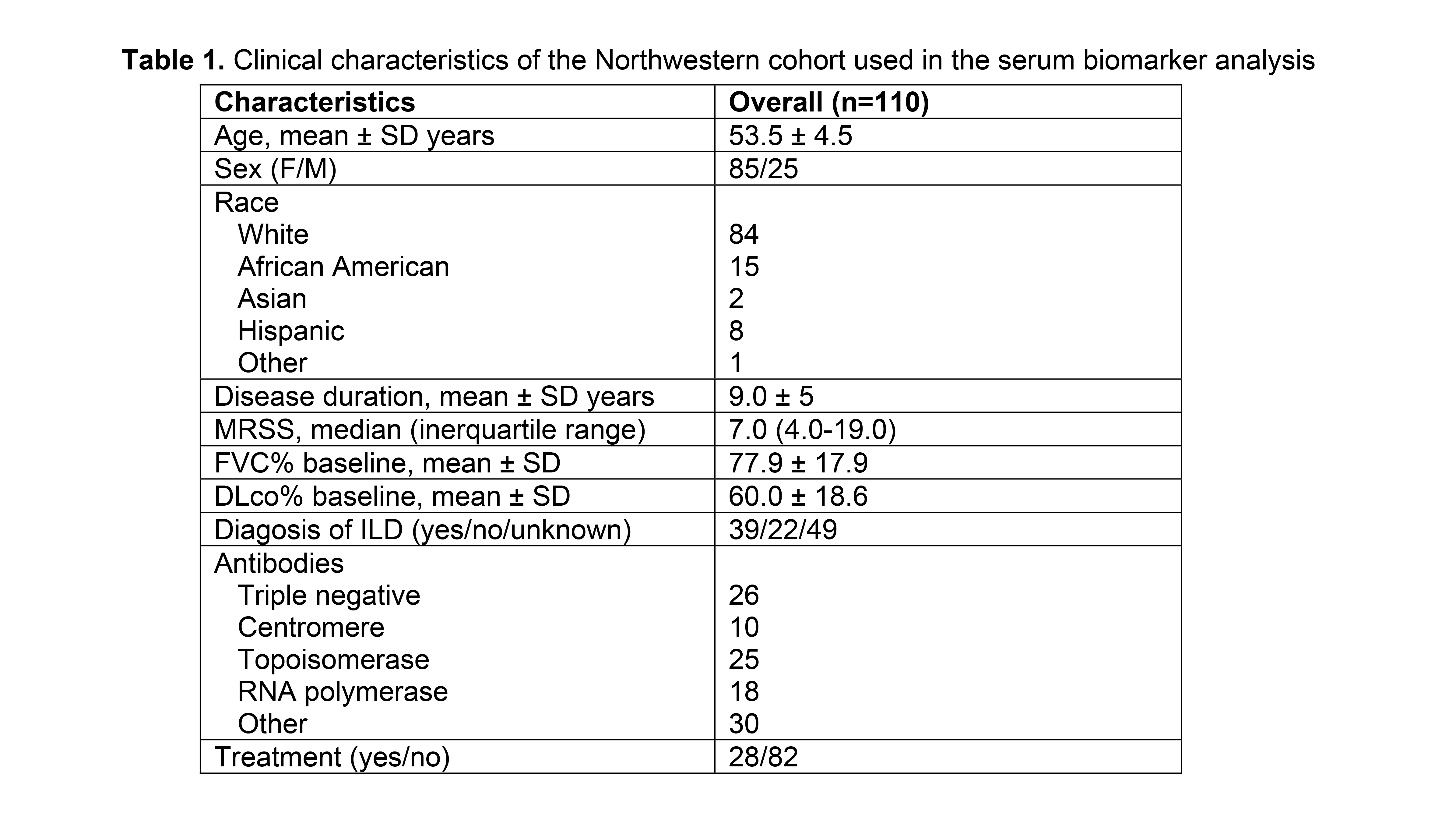
Methods: Our study utilized two patient cohorts for data collection: the Northwestern cohort provided serum samples for biomarker analysis1 while the University of Michigan cohort2 contributed skin samples for scRNA-seq. Senescence markers in the blood (Table 1) were assayed by Luminex xMAP technology. The least absolute shrinkage and selection operator regression was used to select a minimum set of biomarkers to predict individual clinical features followed by logistic regression analysis to determine the discriminatory power of selected biomarkers. The R package Seurat (v4.3.0) was used for analyzing the scRNA-seq data obtained from skin samples (18 controls and 22 SSc patients)2. The Wilcoxon Rank Sum Test is used to compare the difference in cellular senescence score between groups. We considered a P value below 0.05 to be indicative of statistical relevance.
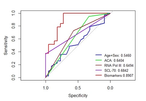
Results: Levels of a combination of 9 putative senescence markers (Eotaxin, IL7, MMP7, RAGE, SOST, ADAMTS13, TARC, GDF15 and MMP-9) were associated with the presence of interstitial lung disease more robustly than anti-Scl-70, RNA polymerase III, or centromere antibodies (Figure 1). Skin biopsies showed notable increases in senescence, defined using the SenMayo gene list3. Dermal endothelial cells, pericytes, and fibroblasts showed the highest SenMayo scores. Among fibroblast subpopulations, a unique SSc-associated COL8A1 cluster showed high senescence score. Moreover, CDKN1A, a hallmark of cellular senescence was substantially elevated in SSc fibroblast subclusters. Ligand-receptor analysis showed senescent SSc fibroblasts interacting with fibroblasts and dendritic cells. The highest senescence scores were associated with early-stage disease and higher skin scores. Across different age groups, individuals with SSc consistently presented with higher senescence scores in the skin than age-matched healthy controls.
Conclusion: These results indicate that circulating levels of putative senescence markers in SSc patients were associated with the presence of lung involvement. SSc skin biopsies show marked upregulation of cellular senescence markers and active signaling by senescent fibroblasts. Subsequent studies will consider senescent cell targeting as a novel strategy for the treatment of this multifaceted condition.
1. Arthritis Care Res 2023; 75(1):152-157
2. Nat Commun 2024;15(1):210
3. Nat Commun 2022;13(1):4827
To cite this abstract in AMA style:
Dey P, Kato H, Huang S, Gudjonsson J, Khanna D, Varga J, Tsou E. Proteomic and Transcriptomic Analysis Reveal Elevated Senescent Signature in Systemic Sclerosis Fibrosis [abstract]. Arthritis Rheumatol. 2024; 76 (suppl 9). https://acrabstracts.org/abstract/proteomic-and-transcriptomic-analysis-reveal-elevated-senescent-signature-in-systemic-sclerosis-fibrosis/. Accessed .« Back to ACR Convergence 2024
ACR Meeting Abstracts - https://acrabstracts.org/abstract/proteomic-and-transcriptomic-analysis-reveal-elevated-senescent-signature-in-systemic-sclerosis-fibrosis/


