ACR Meeting Abstracts

ACR Meeting Abstracts

  • Meetings
    • ACR Convergence 2024
    • ACR Convergence 2023
    • 2023 ACR/ARP PRSYM
    • ACR Convergence 2022
    • ACR Convergence 2021
    • ACR Convergence 2020
    • 2020 ACR/ARP PRSYM
    • 2019 ACR/ARP Annual Meeting
    • 2018-2009 Meetings
    • Download Abstracts
  • Keyword Index
  • Advanced Search
  • Your Favorites
    • Favorites
    • Login
    • View and print all favorites
    • Clear all your favorites
  • ACR Meetings

Abstract Number: 2427

Degranulating PR3+ Myeloid Cells Characterize Proliferative Lupus Nephritis

Alessandra Ida Celia1, Xiaoping Yang2, Hana Minsky2, Silvia Malvica2, Michelle Petri3, the Accelerating Medicines Partnership in RA/SLE4, Avi Rosenberg5 and Andrea Fava2, 1John Hopkins University of Medicine, Rome, Italy, 2Johns Hopkins University, Baltimore, MD, 3Department of Medicine, Division of Rheumatology, Johns Hopkins University School of Medicine, Timonium, MD, 4Multiple, Multiple, 5Department of Pathology, The Johns Hopkins University School of Medicine, Baltimore, MD

Meeting: ACR Convergence 2023

Keywords: Biomarkers, innate immunity, Lupus nephritis, proteomics, Systemic lupus erythematosus (SLE)

  • Tweet
  • Click to email a link to a friend (Opens in new window) Email
  • Click to print (Opens in new window) Print
Session Information

Date: Tuesday, November 14, 2023

Title: Plenary III

Session Type: Plenary Session

Session Time: 11:00AM-12:30PM

Background/Purpose: As part of the Accelerating Medicines Partnership (AMP), we discovered that urinary PR3, a neutrophil degranulation product, is associated with histological activity, indicating the involvement of neutrophil degranulation in proliferative LN, the most aggressive type of LN. Although mature neutrophils with classical polylobate nuclei are rare in LN kidney biopsies, recent evidence suggests that immature myeloid cells undergoing degranulation play a role in the pathogenesis of LN. Our aim is to investigate the presence of PR3+ cells in LN kidney, their association with histopathological features and to define their immunophenotype.

Methods: We performed multiplexed histology using serial immunohistochemistry (sIHC) on LN kidney biopsies to quantify the expression of PR3 and multiple cell lineage markers (20-plex). Image analysis including deconvolution, cell segmentation, glomerular annotation, and quantitative histology was performed using Indica HALO. The analysis was limited to renal cortex.

Results: A total of 11 patients with LN who underwent a clinically indicated kidney biopsy were enrolled: 6 (55%) with pure proliferative LN (ISN/RPS class III or IV) and 5 (45%) with pure membranous LN. PR3+ cells were identified in all biopsies (range 343-7625 per sample) and most of them did not show a polylobate nucleus. The majority of PR3+ cells were in the tubulointersitium (Figure 1A). However, when present, there was a higher density of PR3+ cells in the glomeruli due to their smaller area(Figure 1A-C). PR3+ cell abundance was higher in proliferative LN, especially in the glomeruli (Figure 1A-C). Glomerular PR3+ cell density very strongly correlated with histological activity measured by the NIH Activity Index (Pearson’s r=0.97, p=5*10-5; Figure 1D). PR3+ cells displayed a unilobate nucleus (Figure 2). Most PR3+ cells coexpressed MPO. We identified 2 subsets of PR3+ cells based the coexpression of other lineage markers (Figures 2 and 3). One group coexpressed CD66b, CD11b, CD15, and (variably) CD10 indicating a degranulating phenotype (Figure 3). The other group expressed CD14, CD163, and CD4 indicating a phagocytic phenotype(Figure 3).The was a smaller subset with features of both groups.

Conclusion: PR3+ cells are abundant in LN, increased in proliferative LN and are strongly associated with histological activity thereby characterizing a more aggressive phenotype. This population densely infiltrated the glomeruli emphasizing a potential role in the endothelial pathogenic process. There were two subsets of kidney-infiltrating PR3+ cells characterized by a phagocytic or a degranulating phenotype; both were mononucleated suggesting a monocyte or nonsegmented neutrophil lineage. We previously showed the association between urinary PR3 and histological activity suggesting that intrarenal PR3+ cells are actively degranulating and therefore likely inducing kidney damage. These findings nominate PR3+ cells as a potential therapeutic target. Spatial transcriptomics and proteomic studies are ongoing to define the lineage and function of these cells.

Supporting image 1

Figure1. Clinicopathological associations of intrarenal PR3+ cells. The majority of PR3+ cells were in the tubulointersitium (A). However, accounting for the smaller glomerular area, there was a higher density of PR3+ cells in the glomeruli (B-C). PR3+ cell abundance was higher in proliferative LN, especially in the glomeruli (A-C). Glomerular PR3+ cell density very strongly correlated with histological activity measured by the NIH Activity Index (Pearson’s r=0.97, p=5*10-5; panel D. The analysis was limited to renal cortex.

Supporting image 2

Figure2. Surface markers expression in PR3+ cells.
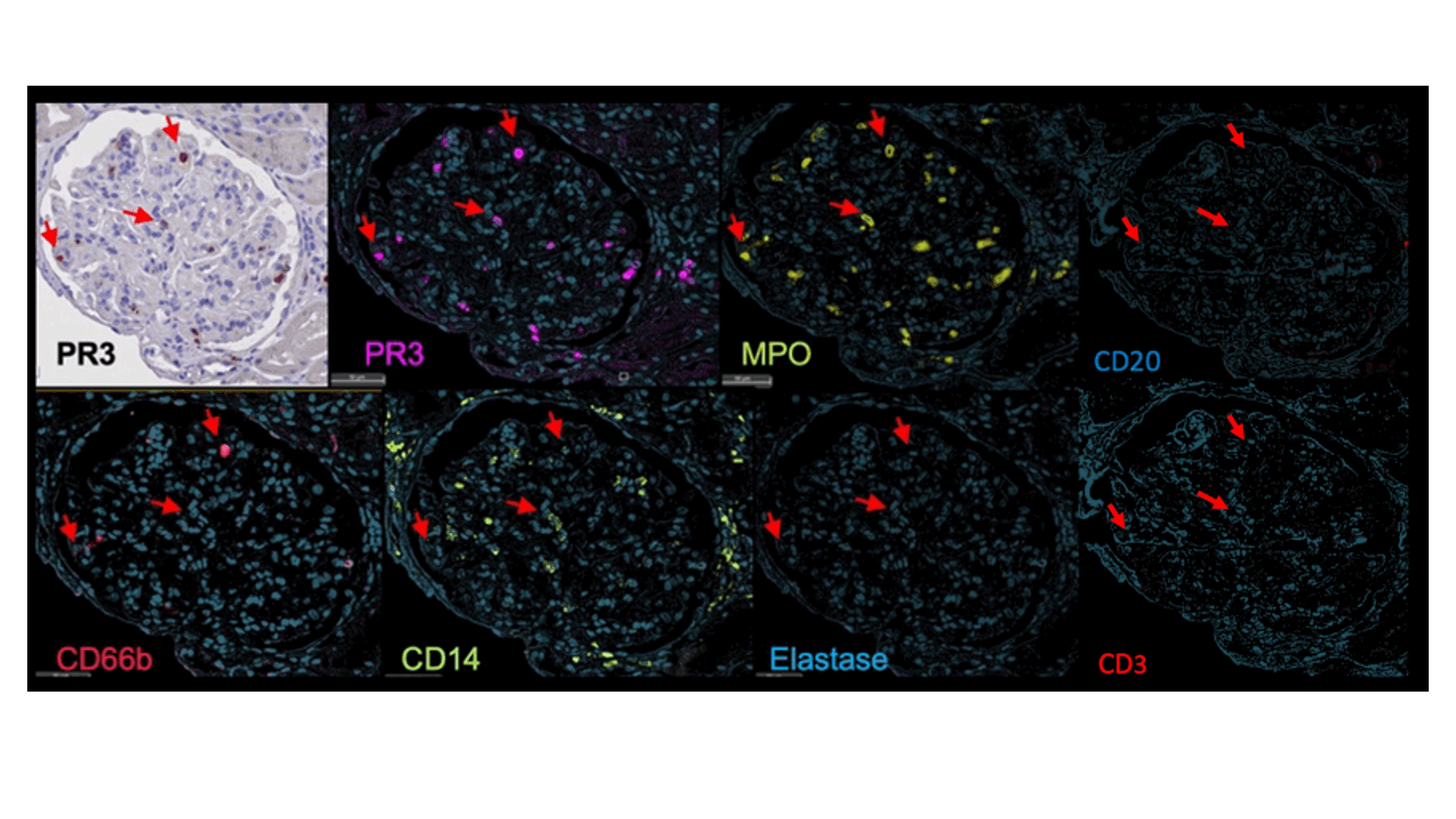
The original and deconvoluted IHC images are shown for a representative glomerulus, with arrows indicating three identical representative cells. Markers indicated at the bottom with matching false colors.

Supporting image 3

Figure3. Cell phenotypes

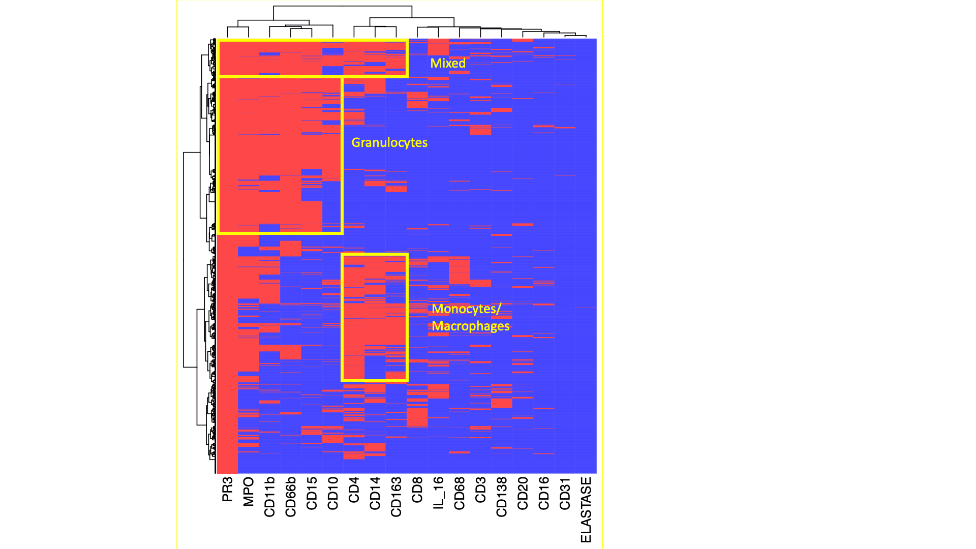
Heatmaps displays the heterogeneity of marker coexpression at the single cell level fo PR3+ cells. Three distinct subsets of PR3+ cells infiltrating the kidney were identified. Data from one representative kidney biopsy (class IV lupus nephritis, NIH Activity Index 8/24, Chronicity Index 6/12).


Disclosures: A. Celia: None; X. Yang: None; H. Minsky: None; S. Malvica: None; M. Petri: Alexion, 1, Amgen, 1, AnaptysBio, 1, Annexon Bio, 1, Argenx, 1, Arhros-Focus Med/Ed, 6, AstraZeneca, 1, 5, Aurinia, 1, 5, 6, Axdev, 1, Biogen, 1, Boxer Capital, 2, Cabaletto Bio, 2, Caribou Biosciences, 2, CVS Health, 1, Eli Lilly, 1, 5, Emergent Biosolutions, 1, Exagen, 5, Exo Therapeutics, 2, Gilead Biosciences, 2, GlaxoSmithKlein(GSK), 1, 5, 6, Horizon Therapeutics, 2, Idorsia Pharmaceuticals, 2, IQVIA, 1, Janssen, 1, 5, Kira Pharmaceuticals, 2, MedShr, 6, Merck/EMD Serono, 1, Momenta Pharmaceuticals, 2, Nexstone Immunology, 2, Nimbus Lakshmi, 2, Proviant, 2, Sanofi, 2, Sinomab Biosciences, 2, Thermofisher, 5, UCB, 2; t. Accelerating Medicines Partnership in RA/SLE: None; A. Rosenberg: None; A. Fava: Annexon Biosciences, 2, Sanofi, 1.

To cite this abstract in AMA style:

Celia A, Yang X, Minsky H, Malvica S, Petri M, Accelerating Medicines Partnership in RA/SLE t, Rosenberg A, Fava A. Degranulating PR3+ Myeloid Cells Characterize Proliferative Lupus Nephritis [abstract]. Arthritis Rheumatol. 2023; 75 (suppl 9). https://acrabstracts.org/abstract/degranulating-pr3-myeloid-cells-characterize-proliferative-lupus-nephritis/. Accessed .
  • Tweet
  • Click to email a link to a friend (Opens in new window) Email
  • Click to print (Opens in new window) Print

« Back to ACR Convergence 2023

ACR Meeting Abstracts - https://acrabstracts.org/abstract/degranulating-pr3-myeloid-cells-characterize-proliferative-lupus-nephritis/

Advanced Search

Your Favorites

You can save and print a list of your favorite abstracts during your browser session by clicking the “Favorite” button at the bottom of any abstract. View your favorites »

All abstracts accepted to ACR Convergence are under media embargo once the ACR has notified presenters of their abstract’s acceptance. They may be presented at other meetings or published as manuscripts after this time but should not be discussed in non-scholarly venues or outlets. The following embargo policies are strictly enforced by the ACR.

Accepted abstracts are made available to the public online in advance of the meeting and are published in a special online supplement of our scientific journal, Arthritis & Rheumatology. Information contained in those abstracts may not be released until the abstracts appear online. In an exception to the media embargo, academic institutions, private organizations, and companies with products whose value may be influenced by information contained in an abstract may issue a press release to coincide with the availability of an ACR abstract on the ACR website. However, the ACR continues to require that information that goes beyond that contained in the abstract (e.g., discussion of the abstract done as part of editorial news coverage) is under media embargo until 10:00 AM ET on November 14, 2024. Journalists with access to embargoed information cannot release articles or editorial news coverage before this time. Editorial news coverage is considered original articles/videos developed by employed journalists to report facts, commentary, and subject matter expert quotes in a narrative form using a variety of sources (e.g., research, announcements, press releases, events, etc.).

Violation of this policy may result in the abstract being withdrawn from the meeting and other measures deemed appropriate. Authors are responsible for notifying colleagues, institutions, communications firms, and all other stakeholders related to the development or promotion of the abstract about this policy. If you have questions about the ACR abstract embargo policy, please contact ACR abstracts staff at [email protected].

Wiley

  • Online Journal
  • Privacy Policy
  • Permissions Policies
  • Cookie Preferences

© Copyright 2025 American College of Rheumatology