Session Information
Date: Sunday, November 12, 2023
Title: (0380–0422) RA – Diagnosis, Manifestations, and Outcomes Poster I
Session Type: Poster Session A
Session Time: 9:00AM-11:00AM
Background/Purpose: While analysis of synovial fluid white blood cell counts (WBCs) is performed in clinical practice for diagnosing septic arthritis and differentiating noninflammatory from inflammatory forms of arthritis, the connection between features of paired synovial fluid and tissue has not been explored. This study seeks to explore the relationship between paired synovial fluid cell counts, synovial histopathology, clinical features, and disease activity.
Methods: Patients meeting ACR/EULAR 1987 and/or 2010 rheumatoid arthritis (RA) classification criteria were recruited prior to elective total knee arthroplasty. All patients provided written informed consent (HSS IRB # 2014-233). Demographics, RA patient reported outcome measures (PROMs), tender and swollen joints, ESR, CRP, RF, and CCP were collected. Synovial fluid was obtained via arthrocentesis performed immediately before surgery. Absolute and differential cell counts were performed, and synovial histopathology using hematoxylin and eosin (H&E) was performed on tissue. An expert musculoskeletal pathologist scored 14 synovial histologic findings: synovial lymphocytic inflammation, mucoid change, fibrosis, fibrin, germinal centers, lining hyperplasia, polymorphonuclear neutrophils (PMNs), detritus, plasma cells, binucleated plasma cells, Russell bodies, sub-lining giant cells, synovial lining giant cells, and mast cells (available at www.hss.edu/pathology-synovitis). Relationships between synovial fluid and tissue data were measured with Pearson’s correlations.
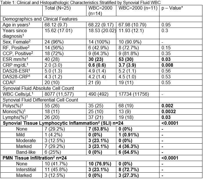
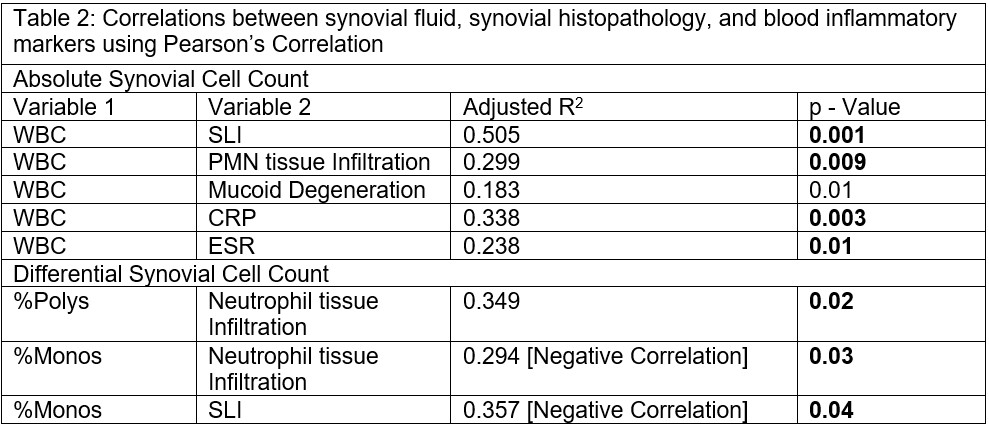
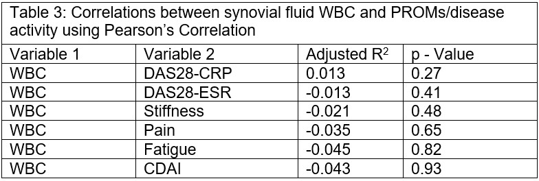
Results: We included 25 patients. Mean age = 68.12 years, mean disease duration = 16.25 years, indicating longstanding disease, mean DAS28-ESR = 4.86 indicating moderate disease activity. Further characteristics of the cohort divided by noninflammatory vs inflammatory synovial fluid (WBC threshold of 2000 cells/microL) are included (Table 1). Synovial fluid WBC was positively correlated with synovial lymphocytic inflammation (SLI), neutrophil infiltration, CRP, and ESR. Of the other synovial histologic features, mucoid degeneration approached significance. Percentage of PMNs in the synovial fluid was positively correlated with PMN infiltration. Percentage of monocytes in the synovial fluid was negatively correlated with PMN infiltration and SLI. Synovial fluid total WBC had no significant correlation with disease activity or PROMs in our cohort. Overview of correlations with associated statistical values included (Table 2, Table 3).
Conclusion: Preliminary results suggest that synovial fluid characteristics are informative and correlate with inflammation in the underlying synovial tissue, but not with disease activity. CRP, present in subclinical synovitis,1 is strongly correlated with synovial fluid WBC. Further analysis of synovial fluid might increase insight into RA pathophysiology and disease control.
References
1. Orange DE, Agius P, DiCarlo EF, et al. Histologic and Transcriptional Evidence of Subclinical Synovial Inflammation in Patients With Rheumatoid Arthritis in Clinical Remission. Arthritis Rheumatol Hoboken NJ. 2019;71(7):1034-1041. doi:10.1002/art.40878
2 = n(%)
*Between groups p-value calculated using t-test.
RF = rheumatoid factor; CCP = cyclic citrullinated protein; ESR = erythrocyte sedimentation rate; CRP = C Reactive Protein; DAS28 = disease activity score using 28 joints; CDAI = clinical disease activity index; WBC = absolute white blood cell count; Polys% = percentage white blood cells in synovial fluid that are polymorphonuclear neutrophils; Monos% = percentage white blood cells in synovial fluid that are monocytes; Lymphs(%) = percentage white blood cells in synovial fluid that are lymphocytes; PMN = polymorphonuclear neutrophils;
Bolded values significant p<0.05
To cite this abstract in AMA style:
Spolaore E, DiCarlo E, Ramirez D, Smith M, Lakhanpal A, Mehta B, Donlin L, Goodman S, Orange D. Synovial Fluid Cell Counts Associated with Joint Histopathology in Rheumatoid Arthritis [abstract]. Arthritis Rheumatol. 2023; 75 (suppl 9). https://acrabstracts.org/abstract/synovial-fluid-cell-counts-associated-with-joint-histopathology-in-rheumatoid-arthritis/. Accessed .« Back to ACR Convergence 2023
ACR Meeting Abstracts - https://acrabstracts.org/abstract/synovial-fluid-cell-counts-associated-with-joint-histopathology-in-rheumatoid-arthritis/


