Session Information
Date: Saturday, November 7, 2020
Title: RA – Diagnosis, Manifestations, & Outcomes Poster II: Biomarkers
Session Type: Poster Session B
Session Time: 9:00AM-11:00AM
Background/Purpose: To better understand site-of-disease mechanisms in arthritis, the phenotypes and organization of synovial cells and infiltrates are critical. Conventional histology-based approaches are limited in the number of proteins that can be detected and quantification can be difficult due to non-linear detection systems. Tissue disaggregation techniques have advantages for some techniques, but disrupt tissue organization, might not liberate all cell types with equal efficiency and can result in loss of cell surface markers. To address this, we applied a novel multiplex immunofluorescence imaging platform (CODEX) to synovial tissue obtained from patients with rheumatoid arthritis (RA) or osteoarthritis (OA).
Methods: CODEX was performed by simultaneously applying 20 different antibodies, each labeled with a unique tag, to a 6 µm frozen tissue section. This technique was followed by iterative marker imaging, data processing and single cell segmentation to generate high resolution images along with marker intensities of each cell. These were used to create a model of cell phenotype, quantification and spatial organization of all identified cell types.
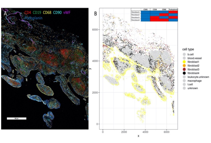
Results: In our initial analysis of 20 markers, we identified the location of multiple subsets, including macrophages, B cells, T cells and mesenchymal cells (Fig 1A). Signals for individual subsets correlated with standard histology in leukocyte-rich and leukocyte-poor tissue sections. One example of how CODEX can identify phenotypically defined cells was fibroblasts, where 4 distinct phenotypes of putative fibroblasts (CD45-) were defined by combinations of podoplanin, CD90, and CD34 (Fig 1B). Type 1 cells (Fb1; podoplanin+ CD34-, CD90-) are almost exclusively found in the intimal lining, whereas Fb2 (CD90+, CD34-, podoplanin-), Fb3 (CD34+, podoplanin+, CD90-), and Fb4 (CD34+, CD90+, podoplanin-) are all located in the sublining. In our small test cohort (4 RA, 3 OA) we noted a nonsigificant numerical increase in total cells in leukocyte-rich RA vs. OA (4994 vs. 2469 average cells/mm2, respectively). The percentage of Fb4 cells was numerically higher in leukocyte-rich RA compared with OA (13.6% vs. 7.5%, respectively).
Conclusion: CODEX technology allows the accurate simultaneous spatial mapping of a multiple cellular phenotypes in situ as suggested here by the location of fibroblast subtypes. Future direction includes combining CODEX with transcriptomics to provide additional characterization of potential pathogenic cells in RA.
 ) Representative CODEX image of RA synovium with CD4 staining in red, CD19 in green, CD68 in yellow, CD90 in cyan, von Willebrand factor (vWF) in magenta and podoplanin in blue. B) Location of 4 distinct phenotypes of putative fibroblasts defined by combinations of podoplanin, CD90, and CD34 expression. Fibroblast type 1 (Fb1) cells are almost exclusively found in the intimal lining, whereas Fb2, Fb3 and Fb4 are all located in the sublining.
To cite this abstract in AMA style:
Wulur I, Boyle D, Zhang L, Martin A, Benschop R, Firestein G. Locating Cellular Subsets in Rheumatoid Arthritis Synovium Using CO-Detection by IndEXing (CODEX) [abstract]. Arthritis Rheumatol. 2020; 72 (suppl 10). https://acrabstracts.org/abstract/locating-cellular-subsets-in-rheumatoid-arthritis-synovium-using-co-detection-by-indexing-codex/. Accessed .« Back to ACR Convergence 2020
ACR Meeting Abstracts - https://acrabstracts.org/abstract/locating-cellular-subsets-in-rheumatoid-arthritis-synovium-using-co-detection-by-indexing-codex/
