ACR Meeting Abstracts

ACR Meeting Abstracts

  • Meetings
    • ACR Convergence 2024
    • ACR Convergence 2023
    • 2023 ACR/ARP PRSYM
    • ACR Convergence 2022
    • ACR Convergence 2021
    • ACR Convergence 2020
    • 2020 ACR/ARP PRSYM
    • 2019 ACR/ARP Annual Meeting
    • 2018-2009 Meetings
    • Download Abstracts
  • Keyword Index
  • Advanced Search
  • Your Favorites
    • Favorites
    • Login
    • View and print all favorites
    • Clear all your favorites
  • ACR Meetings

Abstract Number: 90

SLAMF6 Clustering Is Required to Augment Autoimmune T Cell Activation

Matthew Dragovich1 and Adam Mor 2, 1Columbia University, New York, NY, 2Columbia University School of Medicine, New York, NY

Meeting: 2019 ACR/ARP Annual Meeting

Keywords: Cell Signaling, SLAMF6 and Immunological Synapse, T Cell, TCR

  • Tweet
  • Click to email a link to a friend (Opens in new window) Email
  • Click to print (Opens in new window) Print
Session Information

Date: Sunday, November 10, 2019

Title: T Cell Biology & Targets in Autoimmune & Inflammatory Disease Poster

Session Type: Poster Session (Sunday)

Session Time: 9:00AM-11:00AM

Background/Purpose: The signaling lymphocytic activation molecule (SLAM) family is encompassed by nine distinct receptors that are hematopoietic restricted. Among these, SLAMF6 is highly expressed in autoimmune T cells and genetic variants have been reported in both rheumatoid arthritis and systemic lupus erythematous. Moreover, previous studies as well as our own data, have suggested that anti-SLAMF6 antibodies can have a therapeutic effect in autoimmunity. However, little is known about the role of SLAMF6, specifically in T cell responses. To utilize SLAMF6 interventional approaches, a better understanding of the biology of this receptor in T cell is required. The goal of this work is to uncover SLAMF6 downstream signaling and function in the context of neutralizing antibodies (Abs). We hypothesis that the SLAMF6 is a co-stimulatory receptor in T cells and that blocking its function should disrupt T cell receptor (TCR) signaling, specifically in activated autoimmune T cells.

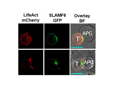
Methods: Primary human T cells were collected from healthy volunteers and from patients with rheumatoid arthritis. Cells were treated with anti-CD3 Abs ± anti-SLAMF6 Abs or anti-CD3 abs ± anti-SLAMF6 monobodies. Cytokine release was measured by ELISA and proliferation was recorded by CFSE dilution assay. SLAMF6 gene was deleted in Jurkat T cells using shRNAs and CRISPR-Cas9, prior to a set of rescue experiments with different versions of mutated SLAMF6 gene. These cells were analyzed by western blot and flow cytometry to illustrate signaling downstream of SLAM6. Confocal microscopy imaging was performed to determine the spatial location of SLAMF6 in the immunological synapse during contact with an antigen presenting cell.

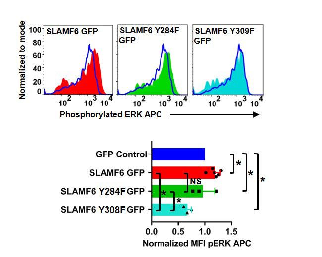
Results: Our experimental data shows that SLAMF6 was obligatory for TCR downstream signaling. Remarkably, SLAMF6 ectodomain was required for its function, yet not for its conscription to the immunological (IS) synapse. Flow-cytometry analysis established that tyrosine 308 of the tail of SLAMF6 was crucial for its ability to augment T cell function (Image 1). Imagining studies revealed that SLAMF6 clustering, with the TCR resulted in a dramatic rise in downstream signaling (Image 2). Mechanistically, we showed that SLAMF6 improved T cell function by increasing T cell adhesivity through activation of the small GTPase Rap1.

Conclusion: We have shown that SLAMF6 ligation via antibodies can deliver a vital co-stimulatory signal to increase TCR activation. This behavior includes increasing cytokine secretion, proliferation,ERK, ZAP70 and AKT phosphorylation and cellular adhesion through Rap1 activation. Our findings propose that during TCR engagement SLAMF6 is transported to the IS independent of its ectodomain where it is engaged by another SLAMF6 molecule through the ectodomain to undergo proper function as a stimulatory co-receptor. SLAMF6 is then deliver a co-stimulatory signal dependent on tyrosine 308 into the T cell. By elucidating the mechanism by which SLAMF6 operates in the TCR pathway, we have provided a model for designing interventional approaches for the treatment of autoimmune diseases where SLAMF6 plays a functional role.

Characteristic histograms of phosphorylated ERK. Jurkat T cells were transfected with GFP control, SLAMF6 GFP, SLAMF6 Y284F GFP and SLAMF6 Y308F GFP then stimulated with CD3 antibodies. After fixation and permeabilization the cells were stained and subjected to flow cytometry. Only GFP positive cells were analyzed for phosphorylated ERK APC. Quantification for a minimum of three independent experiments -n=3-6-. *p<0.05 for an unpaired student t-test. Images and data were adapted from our publication also titled “SLAMF6 Clustering Is Required to Augment T Cell Activation” in press at PLOS ONE -10.1371/journal.pone.0218109-.

Jurkat T cells had been transfected with LifeAct mCherry and SLAMF6 GFP then co-cultured with antigen presenting Raji B cells -APC- -top row- or APCs with SEE -bottom row-. Scale bar is 10μm. Images were adapted from our publication also titled “SLAMF6 Clustering Is Required to Augment T Cell Activation” in press at PLOS ONE -10.1371/journal.pone.0218109-.


Disclosure: M. Dragovich, None; A. Mor, None.

To cite this abstract in AMA style:

Dragovich M, Mor A. SLAMF6 Clustering Is Required to Augment Autoimmune T Cell Activation [abstract]. Arthritis Rheumatol. 2019; 71 (suppl 10). https://acrabstracts.org/abstract/slamf6-clustering-is-required-to-augment-autoimmune-t-cell-activation/. Accessed .
  • Tweet
  • Click to email a link to a friend (Opens in new window) Email
  • Click to print (Opens in new window) Print

« Back to 2019 ACR/ARP Annual Meeting

ACR Meeting Abstracts - https://acrabstracts.org/abstract/slamf6-clustering-is-required-to-augment-autoimmune-t-cell-activation/

Advanced Search

Your Favorites

You can save and print a list of your favorite abstracts during your browser session by clicking the “Favorite” button at the bottom of any abstract. View your favorites »

All abstracts accepted to ACR Convergence are under media embargo once the ACR has notified presenters of their abstract’s acceptance. They may be presented at other meetings or published as manuscripts after this time but should not be discussed in non-scholarly venues or outlets. The following embargo policies are strictly enforced by the ACR.

Accepted abstracts are made available to the public online in advance of the meeting and are published in a special online supplement of our scientific journal, Arthritis & Rheumatology. Information contained in those abstracts may not be released until the abstracts appear online. In an exception to the media embargo, academic institutions, private organizations, and companies with products whose value may be influenced by information contained in an abstract may issue a press release to coincide with the availability of an ACR abstract on the ACR website. However, the ACR continues to require that information that goes beyond that contained in the abstract (e.g., discussion of the abstract done as part of editorial news coverage) is under media embargo until 10:00 AM ET on November 14, 2024. Journalists with access to embargoed information cannot release articles or editorial news coverage before this time. Editorial news coverage is considered original articles/videos developed by employed journalists to report facts, commentary, and subject matter expert quotes in a narrative form using a variety of sources (e.g., research, announcements, press releases, events, etc.).

Violation of this policy may result in the abstract being withdrawn from the meeting and other measures deemed appropriate. Authors are responsible for notifying colleagues, institutions, communications firms, and all other stakeholders related to the development or promotion of the abstract about this policy. If you have questions about the ACR abstract embargo policy, please contact ACR abstracts staff at [email protected].

Wiley

  • Online Journal
  • Privacy Policy
  • Permissions Policies
  • Cookie Preferences

© Copyright 2025 American College of Rheumatology