
Session Information
Date: Sunday, November 17, 2024
Title: Innate Immunity Poster
Session Type: Poster Session B
Session Time: 10:30AM-12:30PM
Background/Purpose: Extracellular vesicles (EVs) are small lipid-bilayer particles released from cells that mediate various functions through their cargo, which includes proteins and nucleic acids. EVs have been linked to the pathogenesis of photosensitive autoimmune diseases such as dermatomyositis (DM) and systemic lupus erythematosus (SLE), and plasma-derived EVs from DM patients have been shown to induce type-1 interferon release. Ultraviolet-B (UVB) radiation can trigger flares of DM and SLE and cause significant skin and systemic inflammation. It is not well-understood how UVB can drive such effects when it is mostly absorbed by the epidermis, with only a small fraction reaching beyond the epidermis. We hypothesize that UVB provokes keratinocytes to release EVs that migrate into the dermis and play a role in dermal and systemic effects.
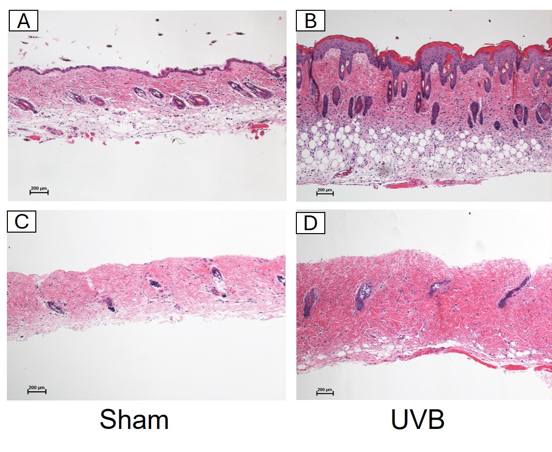
Methods: We irradiated the shaved dorsal skin of 3 male and 3 female C57BL/6 mice for 5 consecutive days with 150 mJ/cm² UVB and used 2 male and 2 female mice as controls. The skin was then harvested and scraped with a blade to remove the epidermis. The dermis was incubated for 30 minutes with a collagenase-dispase enzyme cocktail, and underwent differential ultracentrifugation at 100,000g for 2 hours, followed by iodixanol density-gradient ultracentrifugation at 100,000g for 16 hours and overnight membrane filtration to isolate dermal EVs. Hematoxylin & Eosin and immunohistochemical staining with anti-cytokeratin 1 verified the successful removal of the epidermis (Figure 1).
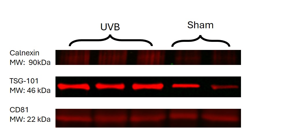
Results: Western blot (WB) and Exoview validated the purity of the EV prep by confirming the expression of tumor-susceptibility gene 101 (TSG101) and the tetraspanin surface marker CD81. Additionally, WB showed minimal expression of Calnexin, an endoplasmic reticulum protein, indicating that the EV prep was nearly free of cellular debris (Figure 2). Electron microscopy showed notable disruption of the basement membrane (BM) of the UVB-irradiated skin compared to the intact BM of sham-irradiated skin. Multiple round vesicle-like structures were visualized near both sides of the BM, more abundantly in the UVB-irradiated skin compared to sham-irradiated skin. Transmission Electron Microscopy of the enriched EV suspension revealed vesicular membrane structures with variable sizes ranging from 20 to 300 nanometers and EV-appropriate morphology.
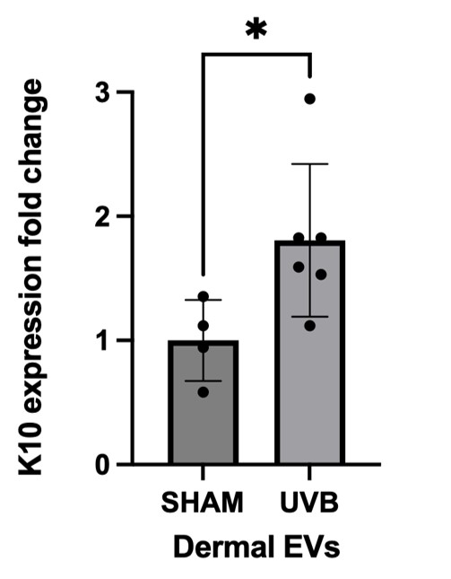
Furthermore, WB, Exoview, and Immunogold electron microscopy (IEM) confirmed the expression of cytokeratin 10 (K10), a marker of keratinocyte differentiation, in the EVs isolated from the dermis, proving that these EVs are indeed epidermally derived from keratinocytes. Moreover, flow cytometry revealed that K10 was significantly more expressed in UVB-irradiated EVs than in sham-irradiated EVs (Figure 3).
Conclusion: These findings suggest that epidermally derived keratinocyte EVs can diffuse through the disrupted basement membrane and reach the dermis, potentially playing a role in the epidermal-dermal crosstalk in UVB-induced dermal and systemic effects seen in photodamage. Further work aims to investigate the immunostimulatory effects of these EVs on dermal and circulating immune cells.
The first and second authors contributed equally to this work.
To cite this abstract in AMA style:
Eldaboush A, Ricco C, Musante L, Dhiman R, Baniel A, Faden D, Stone C, Liu M, Werth V. UVB-Induced Keratinocyte-Derived Extracellular Vesicles: Cytokeratin 10 as a Marker of Epidermal Origin [abstract]. Arthritis Rheumatol. 2024; 76 (suppl 9). https://acrabstracts.org/abstract/uvb-induced-keratinocyte-derived-extracellular-vesicles-cytokeratin-10-as-a-marker-of-epidermal-origin/. Accessed .« Back to ACR Convergence 2024
ACR Meeting Abstracts - https://acrabstracts.org/abstract/uvb-induced-keratinocyte-derived-extracellular-vesicles-cytokeratin-10-as-a-marker-of-epidermal-origin/