ACR Meeting Abstracts

ACR Meeting Abstracts

  • Meetings
    • ACR Convergence 2024
    • ACR Convergence 2023
    • 2023 ACR/ARP PRSYM
    • ACR Convergence 2022
    • ACR Convergence 2021
    • ACR Convergence 2020
    • 2020 ACR/ARP PRSYM
    • 2019 ACR/ARP Annual Meeting
    • 2018-2009 Meetings
    • Download Abstracts
  • Keyword Index
  • Advanced Search
  • Your Favorites
    • Favorites
    • Login
    • View and print all favorites
    • Clear all your favorites
  • ACR Meetings

Abstract Number: 0643

Using FDG PET Brain Scans and Deep Learning Analyses to Identify a Specific Network Correlated with Cognitive Impairment in SLE

Nha Nguyen1, An Vo1, Chris Tang1, Erik Anderson2, Cynthia Aranow2, Betty Diamond3, David Eidelberg1 and Meggan Mackay4, 1Feinstein Institutes, Manhasset, NY, 2Feinstein Institutes for Medical Research, New York, NY, 3The Feinstein Institutes for Medical Research, Manhasset, NY, 4Feinstein Institutes for Medical Research, Manhasset, NY

Meeting: ACR Convergence 2024

Keywords: Biomarkers, Brain, Cognitive dysfunction, Imaging, Systemic lupus erythematosus (SLE)

  • Tweet
  • Click to email a link to a friend (Opens in new window) Email
  • Click to print (Opens in new window) Print
Session Information

Date: Saturday, November 16, 2024

Title: SLE – Diagnosis, Manifestations, & Outcomes Poster I

Session Type: Poster Session A

Session Time: 10:30AM-12:30PM

Background/Purpose: Cognitive impairment (CI) is a debilitating, untreatable problem for up to 80% of individuals with SLE and human and mouse data suggest multiple SLE-related pathogenic mechanisms. However, objective diagnosis of CI with attribution to SLE-mediated mechanisms is difficult, given the limitations of cognitive testing and confounding influences of medications and comorbid disease. We have previously identified hypermetabolic brain regions in SLE subjects that correlate with poor cognitive performance.1 We analyzed FDG PET scans from SLE subjects using deep learning (DL), a form of artificial intelligence, to identify a specific brain network (SLE-CogNet) associated with CI in SLE patients.

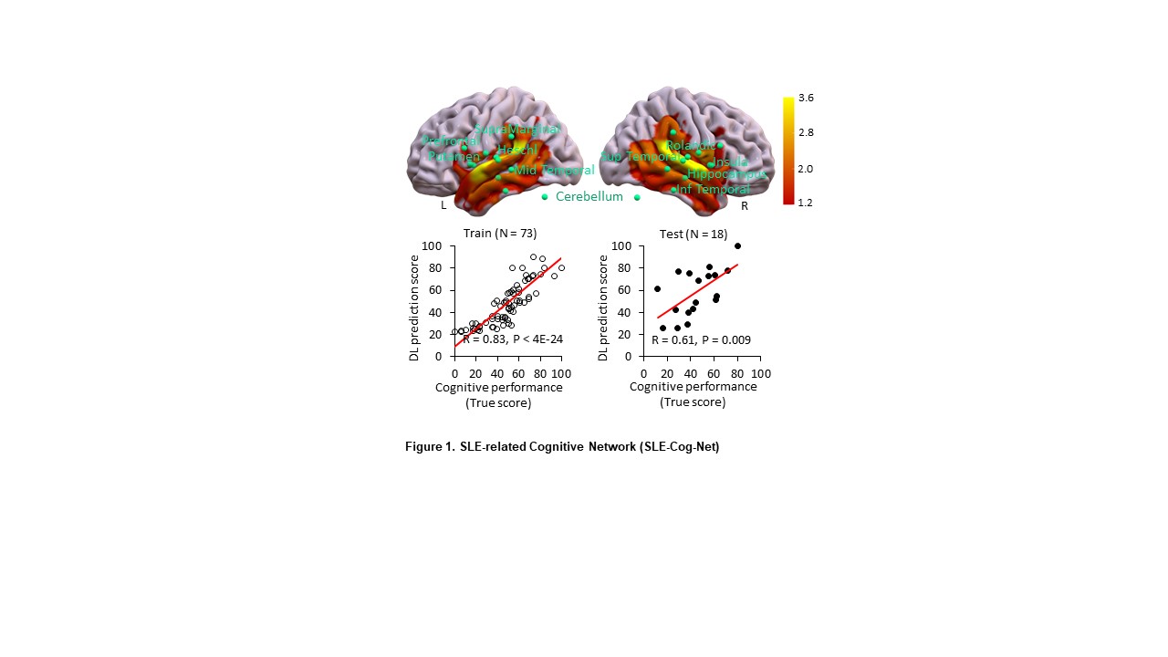
Methods: DL methods were applied to FDG PET data from 91 SLE subjects with concurrent Automated Neuropsychological Assessment Metrics (ANAM) test data. Subjects had stable disease activity, were on prednisone doses ≤ 10 mg, and had no history of CNS disease of any kind. We implemented a 3-dimensional (3D) convolutional neural network (CNN) based on a residual neural network algorithm (ResNet101).2 The cohort was divided randomly into training (n=73: 80%) and testing (n=18: 20%) sets. The 3D CNN was trained on individual FDG PET images and corresponding composite ANAM test scores to identify a significant SLE-CogNet that was validated in the respective testing set. Following training, we employed gradient-weighted class activation mapping (Grad-CAM)3 to generate an explainable map for each scan. DL expression scores for individual SLE subjects were computed across the entire network. For post-hoc analyses, a DL prediction score was determined for 95 regions of interest (ROI) from the automated anatomical atlas as the mean value for the relevant regions.

Results: The resulting SLE-CogNet is characterized by contributions from the hippocampus, prefrontal cortex, putamen, insula, temporal cortex, supramarginal gyrus, rolandic operculum, and cerebellum (Fig. 1). Composite ANAM scores correlated significantly with individual subject DL expression scores for SLE-CogNet in both the training (r=0.83, p< 0.001) and testing (r=0.61, p=0.009) sets. Compared to ANAM data from age and sex matched healthy individuals (n=72), SLE mean ANAM scores were significantly lower (p < 0.01). The R2 measure of the DL model was 69%. Post hoc analyses show significant correlations between C3, anti-dsDNA antibody titers and DL expression scores in multiple regions (p≤0.04).

Conclusion: DL analysis of FDG PET data identifies a candidate SLE-CogNet, composed mainly of hypermetabolic regions associated with CI in previous SLE studies.1 This network may provide an objective measure of CI in SLE that can be used to monitor patients and assess response to treatment.

1 M. Mackay et al., Metabolic and microstructural alterations in the SLE brain correlate with cognitive impairment. JCI Insight 4 (2019)

2 He K et al. Deep Residual Learning for Image Recognition. 2016 IEEE Conference on Computer Vision and Pattern Recognition. (2015)

3 R. Selvaraju et al. Visual Explanations from Deep Networks via Gradient-Based Localization. Int J Comput Vis 128 (2016)

Supporting image 1

Supporting image 2

*AZA (n=8), MTX (n=5), MMF (n=18), Biol (n=17)


Disclosures: N. Nguyen: None; A. Vo: None; C. Tang: None; E. Anderson: Merck/MSD, 3, 11; C. Aranow: AstraZeneca, 1, Bristol-Myers Squibb(BMS), 1, GlaxoSmithKlein(GSK), 5, Kezar Life Sciences Inc, 2, Merck/MSD, 12, Clinical Trial Adjudicator, Synthekine, 1; B. Diamond: adicet, 2, alpine, 12, dsmb, atara, 2, DBV, 2, icell, 2, sail, 2; D. Eidelberg: None; M. Mackay: AbbVie/Abbott, 2, Immunovant, 2, Synthekine, 2, Takeda, 2.

To cite this abstract in AMA style:

Nguyen N, Vo A, Tang C, Anderson E, Aranow C, Diamond B, Eidelberg D, Mackay M. Using FDG PET Brain Scans and Deep Learning Analyses to Identify a Specific Network Correlated with Cognitive Impairment in SLE [abstract]. Arthritis Rheumatol. 2024; 76 (suppl 9). https://acrabstracts.org/abstract/using-fdg-pet-brain-scans-and-deep-learning-analyses-to-identify-a-specific-network-correlated-with-cognitive-impairment-in-sle/. Accessed .
  • Tweet
  • Click to email a link to a friend (Opens in new window) Email
  • Click to print (Opens in new window) Print

« Back to ACR Convergence 2024

ACR Meeting Abstracts - https://acrabstracts.org/abstract/using-fdg-pet-brain-scans-and-deep-learning-analyses-to-identify-a-specific-network-correlated-with-cognitive-impairment-in-sle/

Advanced Search

Your Favorites

You can save and print a list of your favorite abstracts during your browser session by clicking the “Favorite” button at the bottom of any abstract. View your favorites »

All abstracts accepted to ACR Convergence are under media embargo once the ACR has notified presenters of their abstract’s acceptance. They may be presented at other meetings or published as manuscripts after this time but should not be discussed in non-scholarly venues or outlets. The following embargo policies are strictly enforced by the ACR.

Accepted abstracts are made available to the public online in advance of the meeting and are published in a special online supplement of our scientific journal, Arthritis & Rheumatology. Information contained in those abstracts may not be released until the abstracts appear online. In an exception to the media embargo, academic institutions, private organizations, and companies with products whose value may be influenced by information contained in an abstract may issue a press release to coincide with the availability of an ACR abstract on the ACR website. However, the ACR continues to require that information that goes beyond that contained in the abstract (e.g., discussion of the abstract done as part of editorial news coverage) is under media embargo until 10:00 AM ET on November 14, 2024. Journalists with access to embargoed information cannot release articles or editorial news coverage before this time. Editorial news coverage is considered original articles/videos developed by employed journalists to report facts, commentary, and subject matter expert quotes in a narrative form using a variety of sources (e.g., research, announcements, press releases, events, etc.).

Violation of this policy may result in the abstract being withdrawn from the meeting and other measures deemed appropriate. Authors are responsible for notifying colleagues, institutions, communications firms, and all other stakeholders related to the development or promotion of the abstract about this policy. If you have questions about the ACR abstract embargo policy, please contact ACR abstracts staff at [email protected].

Wiley

  • Online Journal
  • Privacy Policy
  • Permissions Policies
  • Cookie Preferences

© Copyright 2025 American College of Rheumatology