Session Information
Date: Tuesday, November 7, 2017
Title: Osteoarthritis – Clinical Aspects Poster II: Observational and Epidemiological Studies
Session Type: ACR Poster Session C
Session Time: 9:00AM-11:00AM
Background/Purpose: Knee osteoarthritis (OA) is more common in women than men; however, the biological mechanisms for sex difference in knee OA are not well understood. Bone shape is a strong risk factor for hip OA, and studies have found a relation with knee OA. The purpose of the present study was to describe knee bone shape changes over time and to examine whether sex is associated with change of bone shape.
Methods: We used data collected from the NIH-funded Osteoarthritis Initiative (OAI), a cohort of persons aged 45-79 at baseline who either had symptomatic knee OA or were at high risk of it. We selected a random sample of 473 knees with radiographs present at baseline, 2 years and 4 years at follow-up visits regardless of OA status. We outlined distal femur and proximal tibia shape on radiographs at all three time points using Statistical Shape Modeling. Group-based trajectory modeling was employed to identify distinctive patterns of bone shape change for each mode. We examined the association between sex and radiographic OA at baseline with trajectories of each bone shape mode using multivariable polytomous regression model while adjusting for age, BMI, race, and results for the first modes for femur and tibia are presented here.
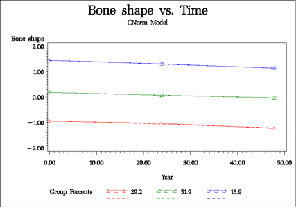
Results: The mean age was 59.9 years (±9.3 SD) for the subjects; the proportion of women was 53.8%. Thirteen modes were derived for proximal tibial shape and for distal femoral shape, accounting for 95.5% of the total variance. In all modes, three distinct trajectory groups were derived with the mean posterior probabilities ranging from 84.3% to 98.8%, indicating excellent model fitting. For the majority of both femoral and tibial modes, the slopes of change were mostly similar within each mode but the intercepts for the 3 trajectory subgroups were different. For femur mode 1, which explained the greatest amount of variance of shape, subgroup mode values decreased slightly over time (see figure); compared with group 1, trajectory subgroup 3 was more likely to include a knee from a woman (OR=28.8, 95% CI: 12.4, 67.1) as was subgroup 2 (OR=4.6, 95%CI: 2.7, 7.9); subgroup 3 was less likely to include knees with OA (OR=0.66; 95%CI 0.56, 0.80), as was subgroup 2 (OR=0.8; 95%CI: 0.7, 0.9). For mode 1 in the tibia, subgroup mode values remained constant over time; compared with group 1, trajectory subgroup 3 was less likely to include womens’ knees (OR=0.6; 95% CI 0.3, 1.1; NS), as was subgroup 2 (OR=0.5; 95%CI 0.3, 0.9); knees with OA were more likely in group 3 (OR=1.3; 95%CI: 1.3, 1.6), but no difference was observed in trajectory membership between groups 2 and 1 for knees with OA.
Conclusion: The shapes of the distal femur and proximal tibia change little over time but do divide into separate trajectory groups largely due to baseline differences that propagate across the years. The trajectory subgroups are associated with both sex and knee OA.
To cite this abstract in AMA style:
Wise BL, Niu J, Zhang Y, Liu F, Pang J, Lynch JA, Lane NE. Trajectories of Knee Bone Shape Change Are Associated with Sex and Osteoarthritis [abstract]. Arthritis Rheumatol. 2017; 69 (suppl 10). https://acrabstracts.org/abstract/trajectories-of-knee-bone-shape-change-are-associated-with-sex-and-osteoarthritis/. Accessed .« Back to 2017 ACR/ARHP Annual Meeting
ACR Meeting Abstracts - https://acrabstracts.org/abstract/trajectories-of-knee-bone-shape-change-are-associated-with-sex-and-osteoarthritis/

