ACR Meeting Abstracts

ACR Meeting Abstracts

  • Meetings
    • ACR Convergence 2024
    • ACR Convergence 2023
    • 2023 ACR/ARP PRSYM
    • ACR Convergence 2022
    • ACR Convergence 2021
    • ACR Convergence 2020
    • 2020 ACR/ARP PRSYM
    • 2019 ACR/ARP Annual Meeting
    • 2018-2009 Meetings
    • Download Abstracts
  • Keyword Index
  • Advanced Search
  • Your Favorites
    • Favorites
    • Login
    • View and print all favorites
    • Clear all your favorites
  • ACR Meetings

Abstract Number: 1977

Three-dimensional Stereophotogrammetry Has Utility in Tracking Upper Eyelid Involvement and Overall Disease Worsening in Patients with Craniofacial Localized Scleroderma

Tyler Cepica1, Jennifer Foster2, Priya Sarlashkar2, Rami Hallac3 and Heidi Jacobe2, 1University of Texas Southwestern Medical School, Benbrook, TX, 2The University of Texas Southwestern Medical Center, Department of Dermatology, Dallas, TX, 3The University of Texas Southwestern Medical Center, Department of Pastic Surgery, Dallas, TX

Meeting: ACR Convergence 2024

Keywords: Cutaneous, Dermatology, Imaging, Outcome measures, Scleroderma

  • Tweet
  • Click to email a link to a friend (Opens in new window) Email
  • Click to print (Opens in new window) Print
Session Information

Date: Monday, November 18, 2024

Title: Imaging of Rheumatic Diseases Poster II

Session Type: Poster Session C

Session Time: 10:30AM-12:30PM

Background/Purpose: Craniofacial localized scleroderma’s (LoS) indolent and subtle course poses challenges in quantifying disease progression. Traditional serial photography and the Localized Scleroderma Cutaneous Assessment Tool (LoSCAT) are validated measures used to assess changes in craniofacial LoS but are limited by their subjective nature and lack of sensitivity to change. Three-dimensional (3D) stereophotogrammetry is a non-invasive imaging strategy with preliminary evidence for utility as an outcome measure in craniofacial LoS, though previous findings have been limited by retrospective design and insufficient comparison to validated measures. As a result, we performed a prospective cohort study assessing changes in craniofacial LoS over time using 3D stereophotogrammetry, serial photography and the LoSCAT. We hypothesized that 3D stereophotogrammetry has greater sensitivity than serial photography and the LoSCAT at detecting changes in craniofacial LoS.

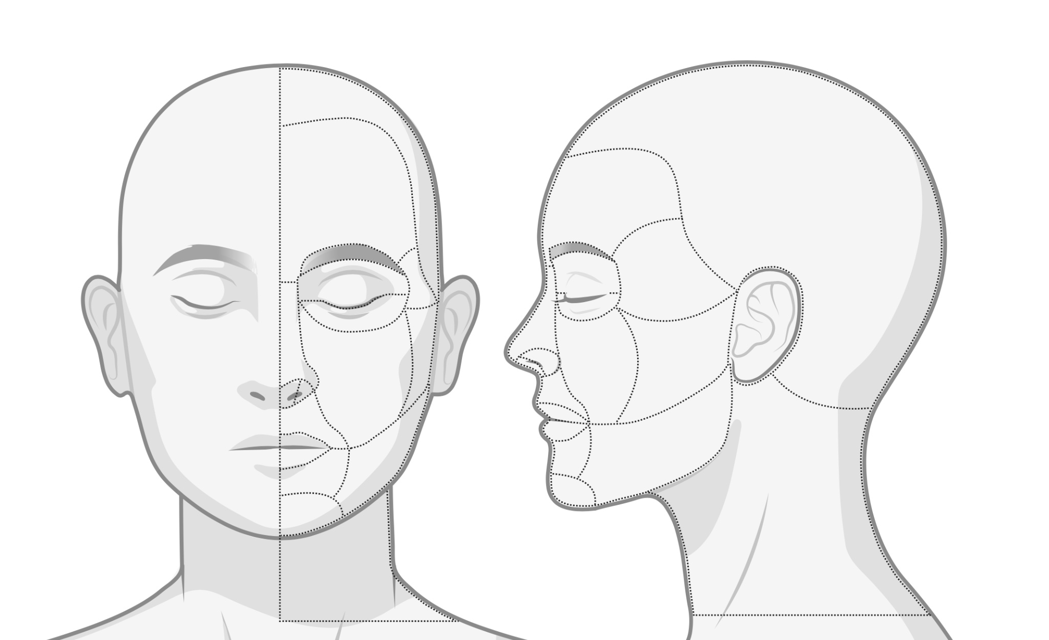
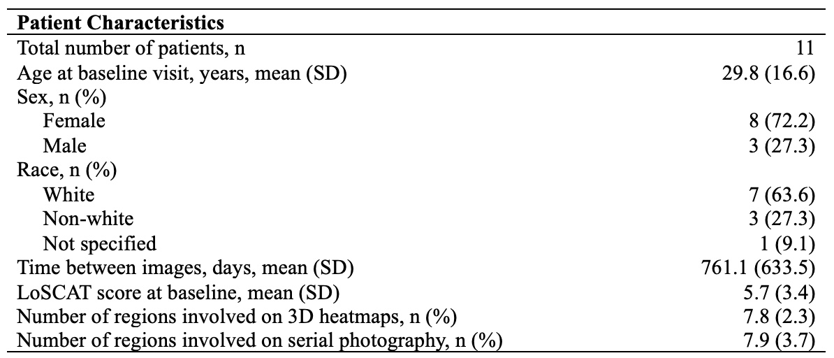
Methods: 11 patients with craniofacial LoS were recruited from an outpatient dermatology clinic and completed two study visits from February 2017 to May 2023. Patients were imaged using standardized protocol in a center dedicated to advanced imaging with 3D and serial photography and had LoSCAT scores assessed by a single expert physician. 3D images were analyzed for asymmetry using in-house mathematical models in MATLAB and converted to 3D heatmaps. Lesion involvement was assessed using 16 distinct craniofacial regions (Figure 1). Worsening and improvement were defined as an increase or decrease in at least one craniofacial region involved or at least one point on the LoSCAT by visit 2. Fisher’s exact and Wilcoxon signed rank tests were performed to compare craniofacial region involvement and patient outcomes for 3D stereophotogrammetry, serial photography, and the LoSCAT.

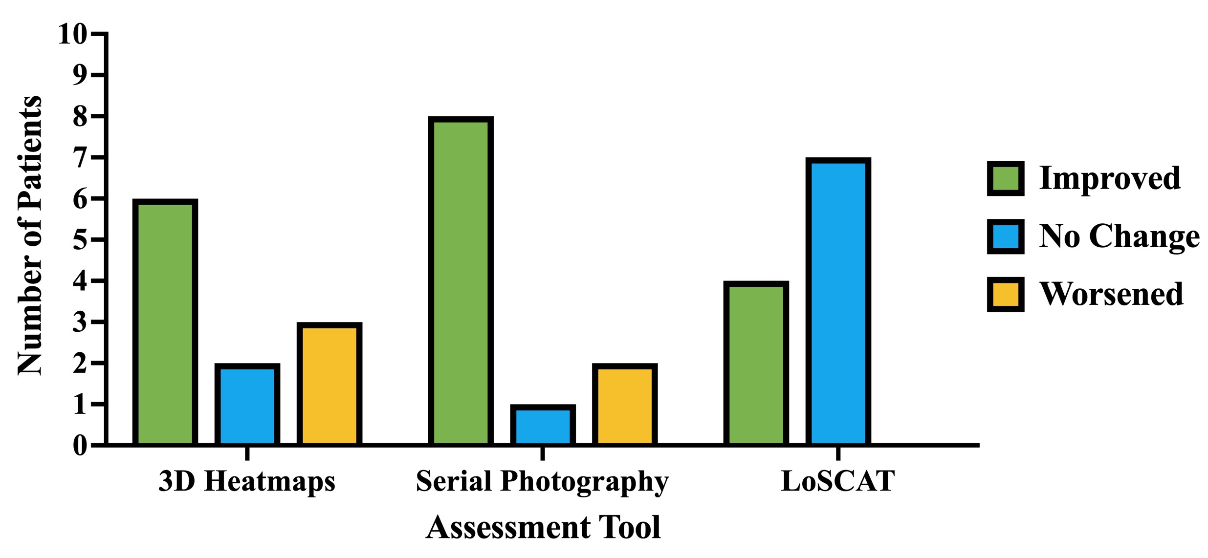
Results: The average number of craniofacial regions involved was 7.8±2.3 for 3D heatmaps and 7.9±3.7 for serial photography (Table 1). Regions most frequently involvement included the lower eyelid and temple (n=17, 77.3%) for 3D heatmaps and the forehead and chin (n=17, 77.3%) for serial photography. 3D heatmaps were more sensitive at detecting involvement of the upper eyelid (p=0.001) while serial photography was more sensitive at detecting involvement of the scalp (p=0.002) and lateral nasal sidewall (p=0.012). Of all measures, the LoSCAT had the highest proportion of patients with no change (n=7, 63.6%), 3D heatmaps had the highest proportion of patients with worsening (n=3, 27.3%), and serial photography had the highest proportion of patients with improvement (n=8, 72.7%) (p=0.040) (Figure 2).

Conclusion: 3D stereophotogrammetry is more sensitive than serial photography at assessing upper eyelid involvement in craniofacial LoS but did not perform as well in assessing involvement of the scalp and lateral nasal sidewall. Importantly, in corroborating previous findings, 3D stereophotogrammetry has greater sensitivity in detecting worsening than either serial photography or the LoSCAT. Furthermore, the existing LoSCAT has poor sensitivity to assess changes in craniofacial LoS, underscoring the need for validation of a craniofacial LoSCAT measure.

Supporting image 1

Sixteen distinct craniofacial regions including: scalp, forehead, brow, upper eyelid, lower eyelid, temple, medial cheek, lateral cheek, lateral nasal sidewall, medial nostril, lateral nostril, upper lip, lower lip, chin, jaw, and neck.

Supporting image 2

Frequency of improved, stable, and worsened disease trends for localized scleroderma regional involvement according to three-dimensional stereophotogrammetry, traditional serial photography, and the LoSCAT. Abbreviations: 3D – three-dimensional; LoSCAT – Localized Scleroderma Cutaneous Assessment Tool.

Supporting image 3

Patient characteristics. Abbreviations: LoSCAT – Localized Scleroderma Cutaneous Assessment Tool; 3D – three-dimensional.


Disclosures: T. Cepica: None; J. Foster: None; P. Sarlashkar: None; R. Hallac: None; H. Jacobe: None.

To cite this abstract in AMA style:

Cepica T, Foster J, Sarlashkar P, Hallac R, Jacobe H. Three-dimensional Stereophotogrammetry Has Utility in Tracking Upper Eyelid Involvement and Overall Disease Worsening in Patients with Craniofacial Localized Scleroderma [abstract]. Arthritis Rheumatol. 2024; 76 (suppl 9). https://acrabstracts.org/abstract/three-dimensional-stereophotogrammetry-has-utility-in-tracking-upper-eyelid-involvement-and-overall-disease-worsening-in-patients-with-craniofacial-localized-scleroderma/. Accessed .
  • Tweet
  • Click to email a link to a friend (Opens in new window) Email
  • Click to print (Opens in new window) Print

« Back to ACR Convergence 2024

ACR Meeting Abstracts - https://acrabstracts.org/abstract/three-dimensional-stereophotogrammetry-has-utility-in-tracking-upper-eyelid-involvement-and-overall-disease-worsening-in-patients-with-craniofacial-localized-scleroderma/

Advanced Search

Your Favorites

You can save and print a list of your favorite abstracts during your browser session by clicking the “Favorite” button at the bottom of any abstract. View your favorites »

All abstracts accepted to ACR Convergence are under media embargo once the ACR has notified presenters of their abstract’s acceptance. They may be presented at other meetings or published as manuscripts after this time but should not be discussed in non-scholarly venues or outlets. The following embargo policies are strictly enforced by the ACR.

Accepted abstracts are made available to the public online in advance of the meeting and are published in a special online supplement of our scientific journal, Arthritis & Rheumatology. Information contained in those abstracts may not be released until the abstracts appear online. In an exception to the media embargo, academic institutions, private organizations, and companies with products whose value may be influenced by information contained in an abstract may issue a press release to coincide with the availability of an ACR abstract on the ACR website. However, the ACR continues to require that information that goes beyond that contained in the abstract (e.g., discussion of the abstract done as part of editorial news coverage) is under media embargo until 10:00 AM ET on November 14, 2024. Journalists with access to embargoed information cannot release articles or editorial news coverage before this time. Editorial news coverage is considered original articles/videos developed by employed journalists to report facts, commentary, and subject matter expert quotes in a narrative form using a variety of sources (e.g., research, announcements, press releases, events, etc.).

Violation of this policy may result in the abstract being withdrawn from the meeting and other measures deemed appropriate. Authors are responsible for notifying colleagues, institutions, communications firms, and all other stakeholders related to the development or promotion of the abstract about this policy. If you have questions about the ACR abstract embargo policy, please contact ACR abstracts staff at [email protected].

Wiley

  • Online Journal
  • Privacy Policy
  • Permissions Policies
  • Cookie Preferences

© Copyright 2025 American College of Rheumatology