Session Information
Session Type: ACR Concurrent Abstract Session
Session Time: 2:30PM-4:00PM
Background/Purpose:
Ultrasound is more sensitive than conventional radiographs in detecting
small osteophytes in hand osteoarthritis (OA). Osteophytes can be seen prior to
joint space narrowing (JSN), may be an early risk factor for OA progression,
and is associated with pain. However, previous studies on the predictive value
of osteophytes on disease progression is limited in numbers, have shown
inconsistent results, and none of these have included ultrasound.
We believe that ultrasound can be used to detect OA at
an earlier stage than conventional radiographs. Hence, the aim of this
longitudinal study was to examine whether ultrasound-detected osteophytes in
joints with concurrent normal radiographs at baseline could predict incident radiographic
hand OA five years later.
Methods: We
included 78 participants (71 women, mean (SD) age 67.8 (5.2) years) from the
Oslo Hand OA cohort. The presence of ultrasound-detected osteophytes was
examined at baseline by two sonographers in collaboration. In the present
analysis we included the joints most likely to develop OA (the first
carpometacarpal joint and the interphalangeal joints bilaterally). One reader
scored the radiographs with known time sequence according to the
Kellgren-Lawrence (KL) scale and the Osteoarthritis Research Society
International (OARSI) atlas for osteophytes and JSN.
Associations between baseline ultrasound-detected
osteophytes (independent variable) and incident radiographic OA features
(dependent variables) were explored only in joints without radiographic OA at
baseline (i.e. KL grade = 0 and no osteophytes or JSN) by use of Generalized
Estimating Equations (GEE), expressed as odds ratio (OR) with 95% confidence
intervals (CI). Analyses were adjusted for age, sex, body mass index and
follow-up time, with significance level 0.05.
Results: Mean
(SD) follow-up time was 4.7 (0.4) years. In total 301 joints were assessed as
being normal on conventional radiographs at baseline, of which 86 had
concurrent osteophytes on ultrasound; most of these were small (score 1 = 79%).
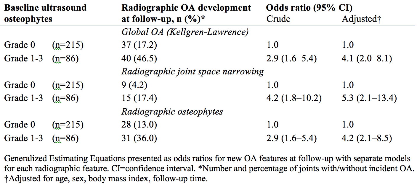
Ultrasound-detected osteophytes at baseline were strong
predictors for incident radiographic OA during follow-up (table). N=40 (47%) of
the joints with preliminary sonographic osteophytes had developed radiographic
OA (i.e. KL score increased from 0) at follow-up, as opposed to only 17% of the
joints without baseline osteophytes. The strongest association was seen for
incident JSN development (OR=5.3, 95% CI 2.1-13.4).
Conclusion: In the
current analysis, we demonstrated that ultrasound-detected osteophytes in
joints assessed as normal on radiographs were a strong predictor for
development of radiographic OA in the same joint five years later. These
results support the use of ultrasound as a promising clinical tool for early
detection of hand OA.
Table
Ultrasound-detected osteophytes at baseline as a
predictor for development of radiographic osteoarthritis (OA)
To cite this abstract in AMA style:
Mathiessen A, Slatkowsky-Christensen B, Kvien TK, Haugen IK, Hammer HB. The Value of Early Ultrasound-Detected Osteophytes in Hand Osteoarthritis: Predicting the Future [abstract]. Arthritis Rheumatol. 2015; 67 (suppl 10). https://acrabstracts.org/abstract/the-value-of-early-ultrasound-detected-osteophytes-in-hand-osteoarthritis-predicting-the-future/. Accessed .« Back to 2015 ACR/ARHP Annual Meeting
ACR Meeting Abstracts - https://acrabstracts.org/abstract/the-value-of-early-ultrasound-detected-osteophytes-in-hand-osteoarthritis-predicting-the-future/

