Session Information
Session Type: Abstract Submissions (ACR)
Background/Purpose
Available studies of craniocervical junction (CCJ) involvement in ankylosing spondylitis (AS) are based on conventional radiography, which has limited ability in the definition of many elements of the CCJ. The goal of the present study was to describe the spectrum of computed tomography (CT) findings in the CCJ in a cohort of patients with AS.
Methods
CT scans of the cervical spine of 11 patients with AS were reviewed and imaging findings related to the CCJ assessed. The standard anatomic intervals describing the CCJ were measured and compared to accepted normal standards. Findings, representing pathology were described, categorized by localization and relation to joints or ligaments of the CCJ.
Results
All patients were males with median age of 48 years and median disease duration of 20 years. The calculated median modified Stoke Ankylosing Spondylitis Spinal Score (mSASSS) for the cervical spine was 8.5, ranging from 0 to 27. Disease-related changes in one or more elements of the CCJ were detected in all patients. Patients with AS had a strong tendency to the narrowing of the atlanto-occipital joints and atlanto-dental interval, with some patients demonstrating complete fusion of these articulations. Atlanto-occipital joints were involved in 8 patients, while 3 patients had disease of the atlanto-dental articulation. Enthesopathy of the CCJ was observed in 7 patients. Significant changes of CCJ were observed also in patients with very low mSASS.
Conclusion
The CCJ is frequently involved in AS patients with advanced disease and may be independent on the mSASSS. Both articulations and ligaments of CCJ may be affected in AS patients.
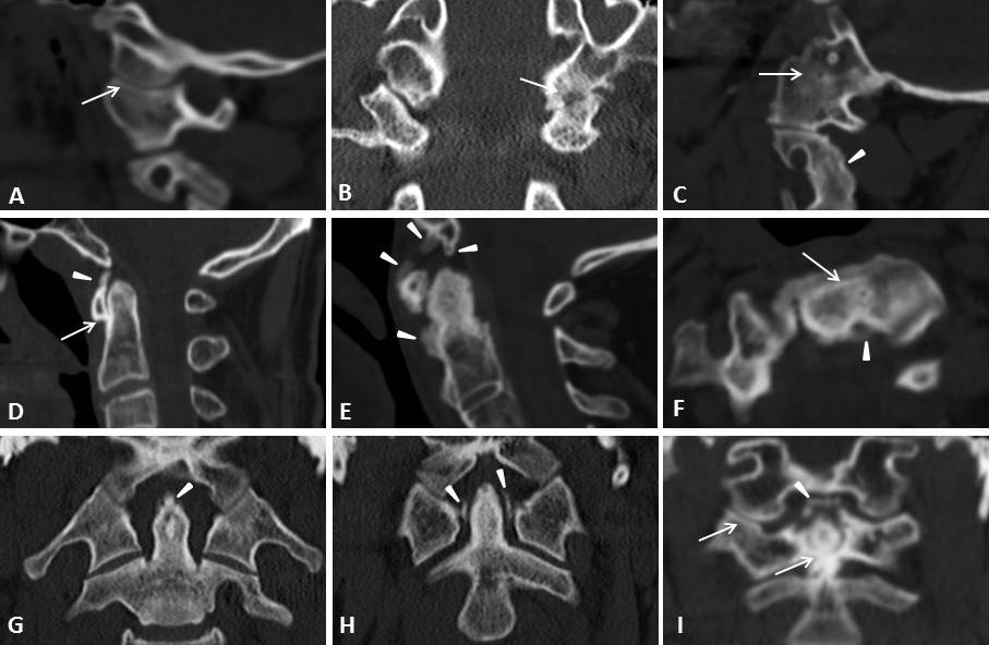
Fig 1. Ankylosing spondylitis of craniocervical junction
- Mild atlanto-occipital joint space narrowing (arrow), saggital view
- Significant narrowing of the left atlanto-occipital joint with erosion (arrow) and osseous sclerosis; right atlanto-occipital joint looks normal
- Total ankylosis of the atlanto-occipital joint (arrow) with secondary osteopenia. Fusion of C2 and C3 parts is also seen (arrowhead)
- Anterior atlanto-dental interval shortening (arrow) and ossification of the anterior atlanto-occipital ligament (arrowhead)
- Severe osseous sclerosis of the dens and C1 arch. Rude proliferative enthesopathy of the anterior longitudinal ligament and ossification of the occipital attachment of the apical ligament (arrowheads)
- Ankylosis of the atlanto-axial joint (arrow), axial view; ossification of the left part of the transverse ligament (arrowhead)
- Apical ligament enthesopathy (arrowhead), coronal view
- Transverse ligament ossification (arrowheads), coronal view
- Alar ligament ossification (arrowhead), coronal view. Narrowing of the right atlanto-occipital and atlanto-axial joints are seen (arrows)
Disclosure:
G. Slobodin,
None;
A. Shpigelman,
None;
H. Dawood,
None;
D. Rimar,
None;
S. Croitoru,
None;
N. Boulman,
None;
M. Rozenbaum,
None;
L. Kaly,
None;
I. Rosner,
None;
M. Odeh,
None.
« Back to 2014 ACR/ARHP Annual Meeting
ACR Meeting Abstracts - https://acrabstracts.org/abstract/the-craniocervical-junction-in-ankylosing-spondylitis-a-computed-tomography-based-study/