Session Information
Session Type: Abstract Submissions (ACR)
Methods: 38 consecutive SSc patients were recruited from SSc Clinic and compared to 51 healthy controls (HC) which were age and body mass index (BMI) matched from the Veteran’s Affair Medical Center Utah Vascular Research Lab. Duplex ultrasonography was utilized to assess baseline vascular parameters forearm blood flow and brachial artery diameter. Afterward a blood pressure cuff was inflated to suprasystolic pressures (240 mmHg) distal to the ultrasound probe on the forearm and brief ischemia was induced for 5 minutes. Following cuff release measure of vascular reactivity (peak reactive hyperemia and hyperemia area under the curve [AUC]), brachial artery FMD (cm and % vasodilation) were obtained. A T-test was used to measure these continuous variables between SSc and HC, as well as SSc patients with DU and without DU.
Results: The SSc population’s mean age was 54±3, with a BMI of 27±1, 33 were female, and 34 were white. Mean mRSS was 8; 31 with limited and 7 with diffuse cutaneous skin distribution. Mean duration of Raynaud’s was 14.9 years and SSc (first non-Raynaud’s symptom) was 11 years. Vasculopathy manifestations included 13 with DU, 6 with pulmonary arterial hypertension, and 1 with scleroderma renal crisis; 31 of these patients were on treatment with vasodilators. The age-matched HC group mean age was 51±2, with a BMI of 24±1 and included 42 females. None of the HC group had a history CVD, diabetes or respiratory disease and were not on prescription medication. None of the subjects in either group used tobacco.
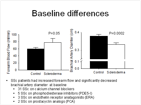
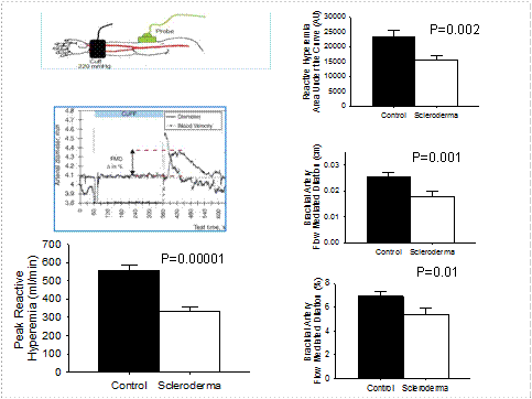
Baseline vascular differences were seen in SSc patients displaying elevated forearm blood flow (p=0.05) and smaller brachial artery diameter (p=0.002) (Figure 1) versus age-matched healthy controls. Post-forearm occlusion, significant differences were seen in hyperemia (p=0.00001), shear stress (p=0.002), brachial artery FMD (cm, p=0.001 and %, p=0.01) compared to healthy controls (Figure 2). Within the SSc population brachial artery flow mediated dilation was significantly different between those with DU and without DU (cm, p=0.05 and %, p=0.05), whereas other parameters were similar (p>0.31).
Conclusion : Utilizing a non-invasive ultrasound technique developed for FMD we identified that SSc patients have significantly altered vascular structure and function which includes: smaller brachial artery diameter, blunted vascular reactivity, attenuated endothelial mediated dilation, despite an increased forearm flow at baseline. SSc patients with DU have a significantly attenuated endothelial mediated dilation, despite having an otherwise similar vascular structure and function profile to those SSc patients without DU. We believe that FMD has potential has clinical utility for identify SSc patients at risk for DU and monitoring response to endothelial-based therapeutics.
Disclosure:
T. M. Frech,
None;
A. Walker,
None;
Z. Barrett-Okeefe,
None;
R. Richardson,
None;
D. W. Wray,
None;
A. Donato,
None.
« Back to 2013 ACR/ARHP Annual Meeting
ACR Meeting Abstracts - https://acrabstracts.org/abstract/the-clinical-utility-of-flow-mediated-dilation-in-systemic-sclerosis/


