Session Information
Date: Monday, November 13, 2023
Title: (1442–1487) SLE – Diagnosis, Manifestations, & Outcomes Poster II
Session Type: Poster Session B
Session Time: 9:00AM-11:00AM
Background/Purpose: Cognitive dysfunction (CD) is highly prevalent in systemic lupus erythematosus (SLE), and advanced structural magnetic resonance imaging (MRI) has revealed brain abnormalities in patients with SLE. We conducted a systematic review of advanced neuroimaging studies to investigate the relationship between structural neuroimaging metrics and CD in SLE.
Methods: A literature search was conducted in accordance with Preferred Reporting Items for Systematic Reviews and Meta-Analyses (PRISMA) guidelines. The search was performed in PUBMED, MEDLINE, EMBASE, Web of Science, and COCHRANE, between January 2000 and April 2022. Both free-text and expanded medical subject headings were used. The search strategy was developed by the research team with the aid of an experienced librarian and included the following terms: systemic lupus erythematosus (SLE), neuropsychiatric lupus (NPSLE), CNS lupus, magnetic resonance imaging (MRI, sMRI), DRI/dMRI, diffusion tensor imaging (DTI), diffusion-weighted imaging (DWI), brain volume, voxel-based morphometry (VBM), cortical thickness, surface area, tractography, fractional anisotropy (FA), mean diffusivity (MD), axial diffusivity (AD), and radial diffusivity (RD). We included observational, case series, cohort, cross-sectional, longitudinal, retrospective, and prospective studies, and selected for neuroimaging studies using MRI and DTI investigating structural brain correlates of cognitive function in SLE.
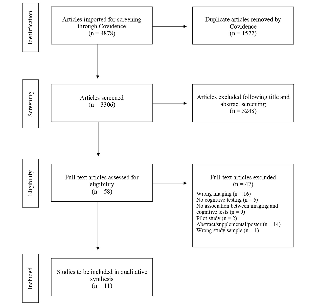
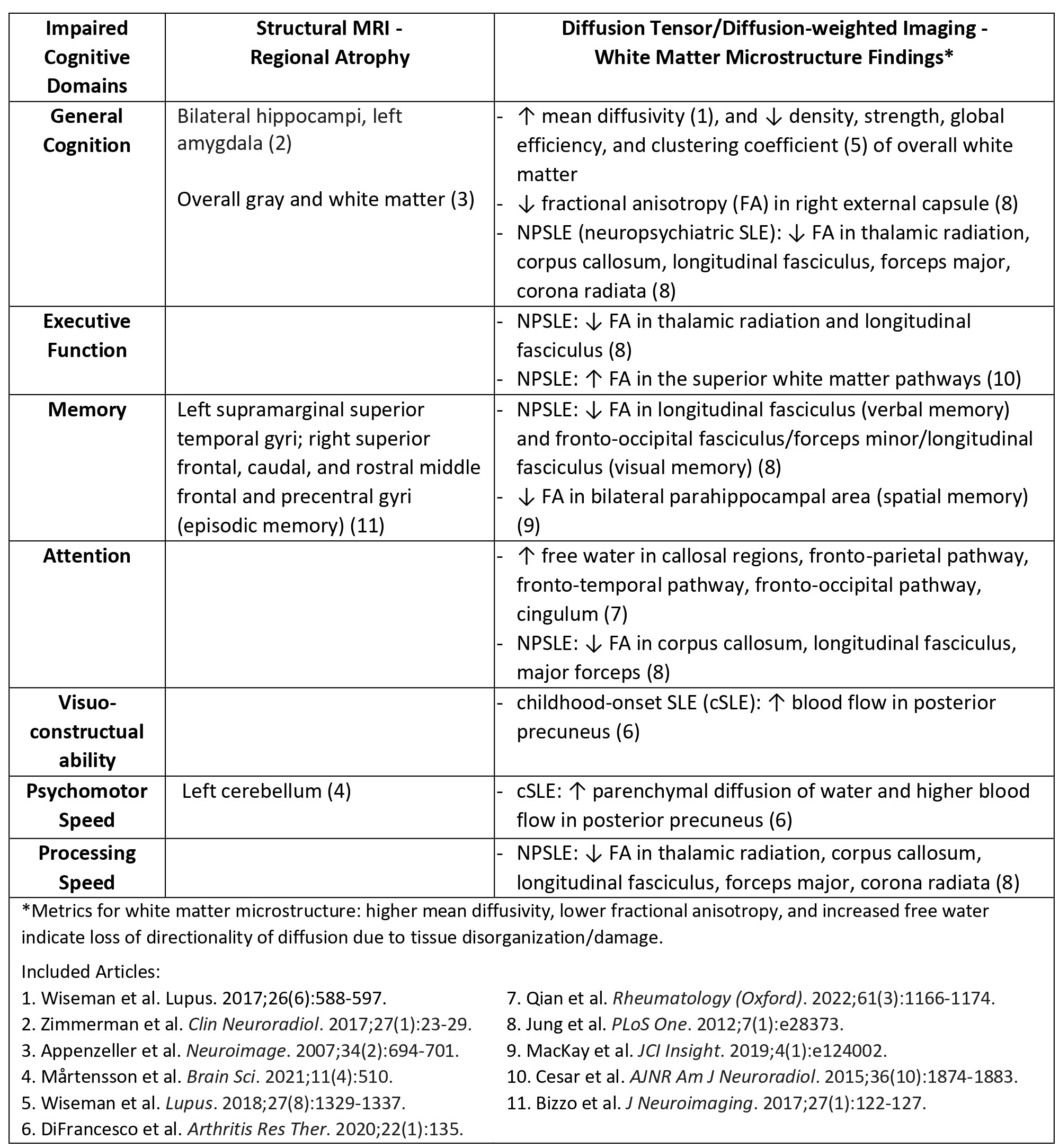
Results: We identified 11 articles meeting search criteria (Figure 1); including two longitudinal studies and only one study in childhood-onset SLE (cSLE). Sample sizes ranged from 22 to 119 (SLE samples, n=11 to 75), and 8 studies included non-SLE controls. Neuroimaging techniques and neurocognitive assessments differed across studies. Structural MRI analysis (n=4 studies) showed cognitive impairment in patients with SLE was associated with overall gray and white matter atrophy, as well as regional atrophy (Table 1). DTI and DWI analyses (n=7 studies) showed associations between cognitive impairment and white matter microstructural changes consistent with tissue disorganization/damage of several regions, in SLE patients with and without NPSLE (Table 1).One longitudinal study found progressive regional atrophy over an average 19-month interval, and the other found stable white matter microstructure over an average 15-month interval.
Conclusion: Studies identified in this systematic review showed abnormalities in brain structure in SLE associated with CD. Limitations included small cross-sectional samples, difficulty in comparing findings across studies due to lack of uniformity in neuroimaging techniques and neurocognitive assessments, and the paucity of pediatric data. Larger, longitudinal studies utilizing standardized measures in both adults and children are needed to better understand the impact of SLE on the brain structure and function across the age spectrum.
To cite this abstract in AMA style:
Mohamed I, El Tal T, arciniegas s, Fevrier S, ledochowski j, Knight A. Systematic Review of Effects of Systemic Lupus Erythematosus on Brain Structure and Structural Connectivity and Its Relationship with Cognitive Dysfunction Through an Advanced Neuroimaging Lens [abstract]. Arthritis Rheumatol. 2023; 75 (suppl 9). https://acrabstracts.org/abstract/systematic-review-of-effects-of-systemic-lupus-erythematosus-on-brain-structure-and-structural-connectivity-and-its-relationship-with-cognitive-dysfunction-through-an-advanced-neuroimaging-lens/. Accessed .« Back to ACR Convergence 2023
ACR Meeting Abstracts - https://acrabstracts.org/abstract/systematic-review-of-effects-of-systemic-lupus-erythematosus-on-brain-structure-and-structural-connectivity-and-its-relationship-with-cognitive-dysfunction-through-an-advanced-neuroimaging-lens/


