Session Information
Session Type: Poster Session A
Session Time: 9:00AM-11:00AM
Background/Purpose: Right and Moll recognized the presence of spinal inflammation/axial disease in psoriatic arthritis (PsA) in their seminal work in 1973. The prevalence of axial disease in patients with PsA varies with disease duration. The Total Body (TB)-PET/CT with the 18F-FDG radiotracer captures glucose metabolism across the entire body, and provides standardized measures as surrogates for degree of inflammation such as SUVmax. The objective of this study was to apply high-sensitivity TB 18F-FDG PET/CT imaging to identify and characterize the spectrum of spinal inflammation in patients with PsA.
Methods: We prospectively recruited 25 PsA patients (6 female, 19 male; mean age of 51.4±16.4 years), as per CASPAR criteria. Among them, 8 patients had inflammatory low back pain (LBP). All subjects underwent a single-time point 18F-FDG TB-PET/CT using 1/5th the standard radioisotope dose (78.2±4.9 MBq) (Abdelhafez Y,et al .J Nucl Med. 2022(10):1579-1585).
Degree of inflammation was quantified by using maximum standardized uptake value ratio corrected by the background level (rSUVmax).Qualitative changes in the joints (atlantoaxial, apophyseal, costovertebral/costotransverse, and sacroiliac) and entheses (cervical/thoracic/lumbar supra- and interspinous ligaments) were also evaluated.
Results: In one or more of the described sites PET imaging demonstrated abnormality in 21 (84%) patients. Notable changes respectively were observed in atlantoaxial (Figure 1), apophyseal, costovertebral/ costotransverse, and sacroiliac joints (Figure 2) in 15 (60%), 18 (72%), 11 (44%), and 5 (20%) patients in variable combinations.
20% patients(5/25) demonstrated sacroiliitis by PET imaging, among them the involvement was unilateral in three and bilateral but asymmetric in two patients (Figure 2). A patient with PsA with inflammatory LBP for >5 years had no evidence of sacroiliitis on MRI but demonstrated unilateral pronounced sacroiliitis on PET.
Increased 18F-FDG uptake suggestive of active inflammation on PET imaging was observed in cervical, thoracic, and lumbar spine enthuses (Figure 3) in 14 (56%), 17 (68%), and 15 (60%) patients, respectively. Overall, spinal enthesitis was evident in 80% (20/25) patients.
Quantification of the total spinal inflammatory load (rSUVmax ) could be measured in each patient, the summed rSUVmax/patient was on average 7.3±3.1 for all the described sites.
Conclusion: TB-PET/CT scan is an US FDA approved technology. We observed spinal inflammation to be a dominant pathology in PsA : i. Our findings provide direct in-vivo evidence that enthesitis could be the primary pathology for axial inflammation in PsA; spinal enthesitis was observed in 80% of patients. In majority of the patients this pathology was clinically occult, as only about one third of the patients reported back pain. ii. Non-radiographic axial disease in PsA remains unaddressed. Here we have demonstrated this condition in PsA and we provide TB-PET/CT as a biomarker tool to diagnose this condition. iii. In-vivo TB-PET imaging provided the total inflammatory burden (rSUVmax) of the whole spine thus demonstrating the ability of TB-PET/CT as a quantitative tool for assessing disease severity and for therapeutic response.
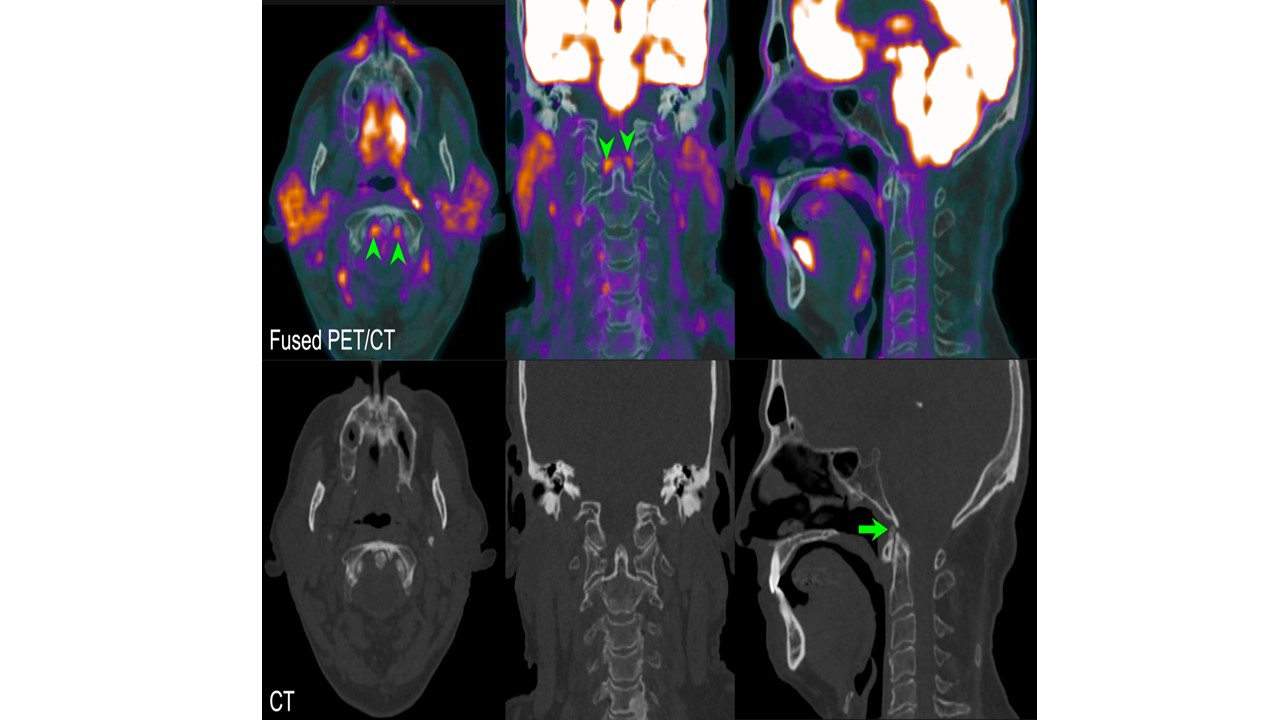
Fused 18F-FDG PET/CT sections (top row) and their corresponding low-dose CT (bottom row) extracted from TB-PET/CT for a 62-year-old man with PsA in axial (left), coronal (middle) and sagittal (right) views. Images demonstrate intense FDG uptake with rSUVmax 1.7 at the alar ligaments (arrowheads) with evidence of ossification of the anterior atlanto-occipital membrane on the CT (arrow).
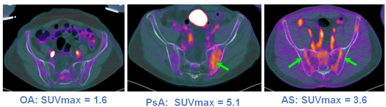
PET-CT imaging is capable of Identifying and providing in vivo quantitative measures of the degree of inflammation (SUVmax) as shown in this figure. Compared to osteoarthritis (OA) marked unilateral SI joint inflammation can be been in psoriatic arthritis (PsA) and bilateral symmetric inflammation of SI joints in ankylosing spondylitis (AS).
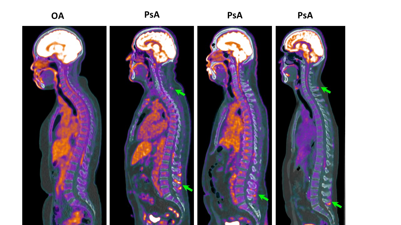
Enthesitis at multiple levels in the spine (green arrows) with or without sacroiliitis is an interesting finding in the psoriatic arthritis (PsA) patients by total body the PET-CT imaging. Where as in osteoarthritis (OA) spinal inflammation could not be detected. Involvement of the inter-/supra-spinous ligament are marked by the green arrows.
To cite this abstract in AMA style:
Raychaudhuri S, Abdelhafez Y, Mazza D, Raychaudhuri S, Chaudhari A. Spinal Inflammation a Dominant Pathology in Psoriatic Arthritis: Characterization and Quantification by In-Vivo 18F-FDG Total-Body PET/CT Imaging [abstract]. Arthritis Rheumatol. 2023; 75 (suppl 9). https://acrabstracts.org/abstract/spinal-inflammation-a-dominant-pathology-in-psoriatic-arthritis-characterization-and-quantification-by-in-vivo-18f-fdg-total-body-pet-ct-imaging/. Accessed .« Back to ACR Convergence 2023
ACR Meeting Abstracts - https://acrabstracts.org/abstract/spinal-inflammation-a-dominant-pathology-in-psoriatic-arthritis-characterization-and-quantification-by-in-vivo-18f-fdg-total-body-pet-ct-imaging/