ACR Meeting Abstracts

ACR Meeting Abstracts

  • Meetings
    • ACR Convergence 2024
    • ACR Convergence 2023
    • 2023 ACR/ARP PRSYM
    • ACR Convergence 2022
    • ACR Convergence 2021
    • ACR Convergence 2020
    • 2020 ACR/ARP PRSYM
    • 2019 ACR/ARP Annual Meeting
    • 2018-2009 Meetings
    • Download Abstracts
  • Keyword Index
  • Advanced Search
  • Your Favorites
    • Favorites
    • Login
    • View and print all favorites
    • Clear all your favorites
  • ACR Meetings

Abstract Number: 1643

Single Nuclei Multiome and Spatial Transcriptomic Analysis of Early, Untreated SSc Skin Identifies Signaling Interactions Between Macrophages and Fibroblasts

Helen Jarnagin1, Dillon Popovich2, Rezvan Parvizi3, Rosemary Gedert4, Lam C. Tsoi5, Rachael Wasikowski5, Zhiyun Gong1, Madeline Morrisson6, Laurent Perreard7, Fred Kolling IV7, Dinesh Khanna4, Johann Gudjonsson4 and Michael Whitfield3, 1Dartmouth College, Lebanon, NH, 2Dartmouth College, West Lebanon, NH, 3Geisel School of Medicine at Dartmouth, Hanover, NH, 4University of Michigan, Ann Arbor, MI, 5Michigan, Dept. of Dermatology, Ann Arbor, MI, 6Geisel School of Medicine at Dartmouth College, Hanover, NH, 7Geisel School of Medicine, Dartmouth College, Lebanon

Meeting: ACR Convergence 2024

Keywords: Fibroblasts, Dermal, Gene Expression, macrophages, skin, Systemic sclerosis

  • Tweet
  • Email
  • Print
Session Information

Date: Sunday, November 17, 2024

Title: Plenary II

Session Type: Plenary Session

Session Time: 9:00AM-10:30AM

Background/Purpose: We generated a vertically integrated dataset on treatment naïve patients with dcSSc (diffuse Systemic Sclerosis) skin that includes bulk RNA-seq, single nuclei multiome, and paired spatial transcriptomic analyses to comprehensively describe the molecular changes in these individuals. Given the well-established immune-fibroblast axis in SSc, we sought to understand the dynamics between macrophages and fibroblasts to identify key pathways driving the disease.

Methods: Skin biopsies and paired PBMC samples were collected from 10 treatment-naïve individuals diagnosed with dcSSc, along with 4 age- and sex- matched healthy controls (HC). Multiple biopsies were taken: one for bulk RNA-seq analysis and molecular subtype assignment, another for single nuclear RNA-seq and ATAC-seq (snMultiome), and the third for spatial transcriptomics. 10X Genomics platforms were used for sequencing, including 10X Visium. Data were analyzed in R, using packages such as Signac, Seurat, PRECAST, CARD, and CellChat. SSc-derived self-assembled (SA) 3D skin-like tissue models were used to test response to therapeutics in vitro.

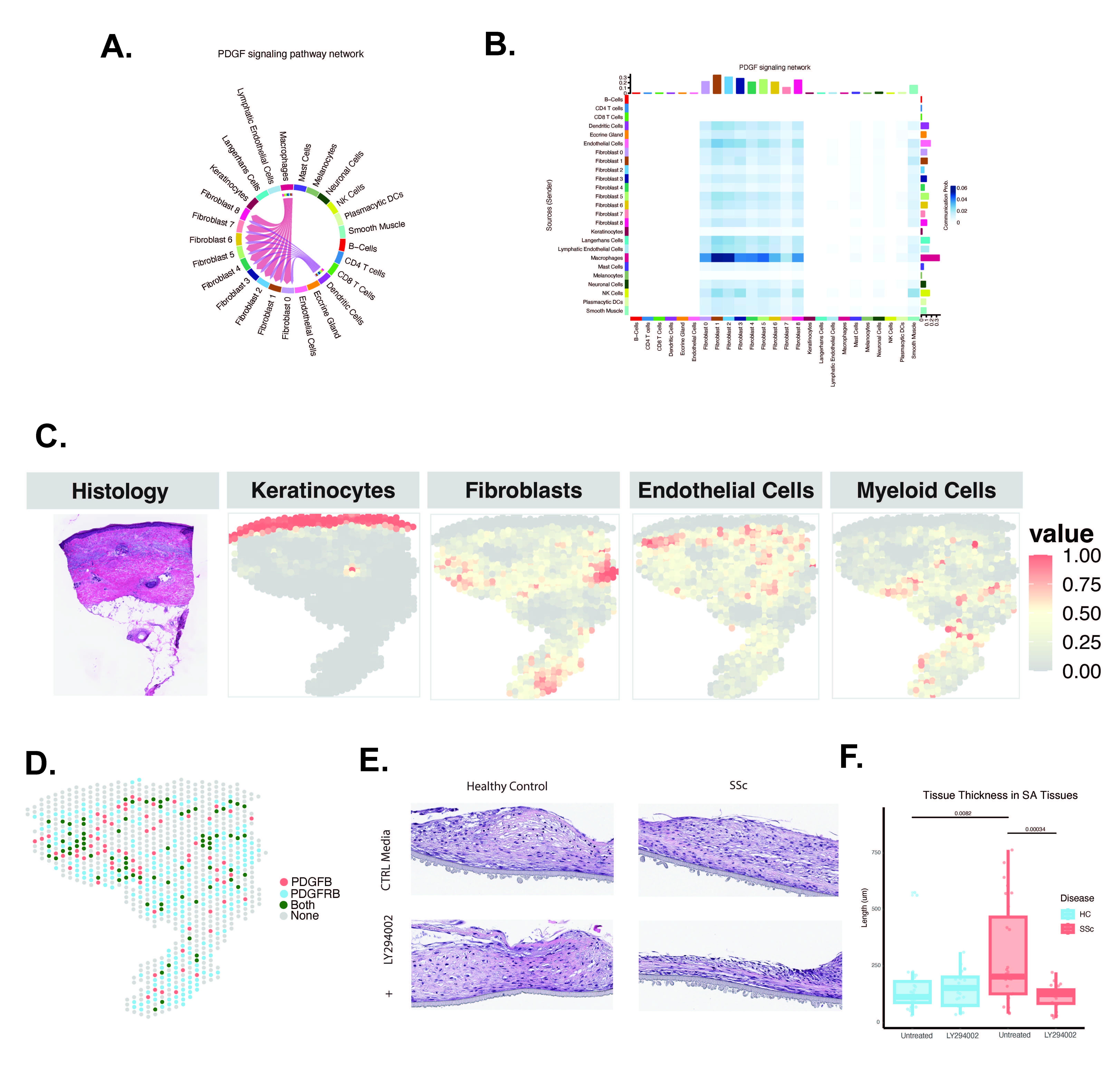
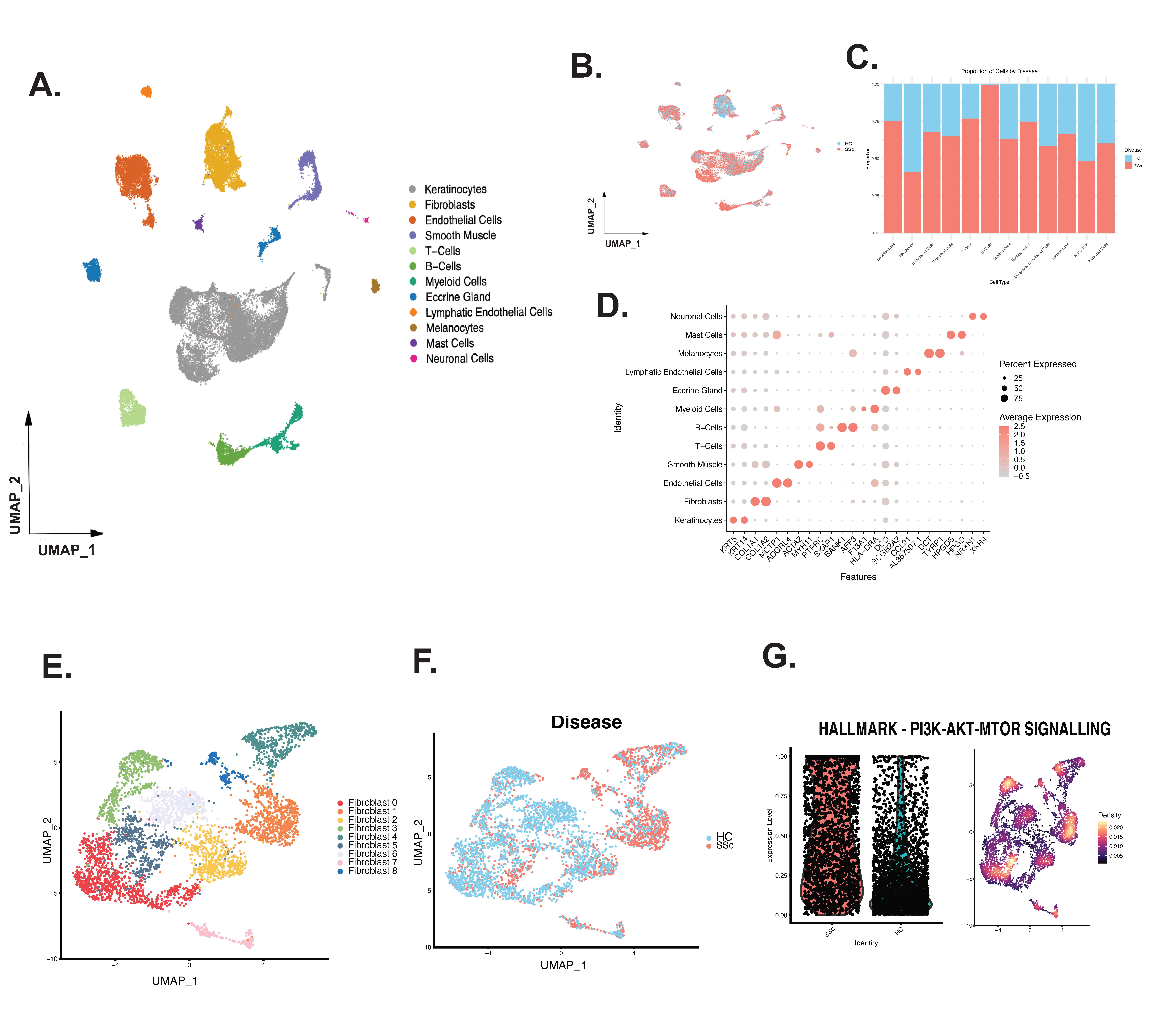
Results: We integrated multiple data types in a cohort of 10 individuals with untreated, early-stage dcSSc. SnMultiome was performed on all cell types; cell clustering showed major population shifts for B-cells and T-cells in SSc samples (Fig 1 A-D). Fibroblast subclustering and pathway analyses showed that SSc patient fibroblasts exhibited enriched PI3K-AKT-mTOR pathway expression largely driven by PDGF and other growth factor signaling (Fig 1 E-G). Transcription factors specific to the PI3K pathway, SRF and Stat5a, were enriched in inflammatory, SSc-dominated fibroblast clusters characterized by high expression of C7, PLA2G2A, CXCL12, and APOE. Ligand-receptor analysis identified signaling pathways between immune cells and fibroblasts. Macrophages had the highest probability of secreting PDGF to interact with the inflammatory SSc-dominant fibroblast subclusters (Fig 2 A-B). Spatial transcriptomic analyses showed that samples with high myeloid cell infiltration had increased PDGF secretion by innate immune cells, matching the increased expression of PDGFR in fibroblasts (Fig 2 C-D). Self-assembled 3D skin-like tissues constructed from SSc fibroblasts treated with a PI3K inhibitor (LY294002) showed significantly reduced tissue thickness post-treatment (Fig 2 E-F). In contrast, tissues constructed from healthy control cells produced thinner tissues and did not show a significant reduction in thickness after treatment with the PI3K inhibitor.

Conclusion: Vertical integration of multiomic data provides unique insights into the molecular interactions between cell types in early, untreated SSc skin. Increased PDGF and downstream PI3K activity is a hallmark of SSc skin fibrosis. PI3K inhibition reduces tissue thickness, providing a specific druggable target for disrupting the immune-fibrosis axis in SSc skin.

Supporting image 1

Figure 1. Complete snMultiome clustering and fibroblast subclustering shows a clear transcriptomic shift in SSc. A) Uniform Manifold Approximation and Projection (UMAP) depicting multi-modal clustering of all nuclei harvested from 10 SSc and 4 HC skin samples. B) UMAP demonstrating broad distribution of SSc and HC nuclei. C) Proportional difference of each cell type by disease status. D) Two of the most differentially expressed RNA transcripts from each cell type were identified in the UMAP. E) UMAP clustering identifies 9 distinct Fibroblast subclusters. F) UMAP demonstrating the broad distribution of SSc and HC nuclei in the subsetted fibroblasts. G) Variance-adjusted Mahalanobis (VAM) analysis reveals significant PI3K Hallmark pathway enrichment in SSc subclusters and SSc fibroblasts compared to HC.

Supporting image 2

Figure 2. Ligand-receptor analysis demonstrates the enrichment of PDGF signaling between Macrophages in Fibroblasts. A) A chord diagram shows the direct signaling of all PDGF ligands from Macrophages to all fibroblast subclusters, with stronger signaling probability than Dendritic Cells to fibroblast subclusters. B) Heatmap shows PDGF ligand-receptor interactions in all cells, including the immune and fibroblast subclusters. The highest probability of PDGF signaling interactions is observed between macrophages and all fibroblast subclusters. C) Representative histology and Visium deconvoluted spots (using CARD) for major cell types in the skin section. D) Visium spot section showing significant expression of PDGFB and PDGFRB, and co-expression in section with high myeloid cell infiltration. E) Representative tissue sections of self-assembled (SA) tissues from HC fibroblasts or SSc fibroblasts either untreated or treated with PI3K inhibitor, LY294002. F) The average difference between SA tissue thickness shows a significant difference in SSc-derived SA tissues treated with LY294002 and HC (p-value 0.00034).


Disclosures: H. Jarnagin: None; D. Popovich: None; R. Parvizi: None; R. Gedert: None; L. Tsoi: Janssen, 5; R. Wasikowski: None; Z. Gong: None; M. Morrisson: None; L. Perreard: Masimo Inc, 5; F. Kolling IV: None; D. Khanna: AbbVie/Abbott, 2, Amgen, 2, AstraZeneca, 2, Boehringer-Ingelheim, 2, Bristol-Myers Squibb(BMS), 2, Cabaletta, 2, Certa Therapeutics, 2, GlaxoSmithKlein(GSK), 2, Janssen, 2, MDI Therapeutics, 8, Merck/MSD, 2, Novartis, 2, Zura Bio, 2; J. Gudjonsson: None; M. Whitfield: Abbvie, 6, Boehringer Ingelheim, 1, 2, Bristol-Myers Squibb, 2, 5, Celdara Medical, LLC, 5, 8, 9, 10, UCB Biopharma, 2, 5.

To cite this abstract in AMA style:

Jarnagin H, Popovich D, Parvizi R, Gedert R, Tsoi L, Wasikowski R, Gong Z, Morrisson M, Perreard L, Kolling IV F, Khanna D, Gudjonsson J, Whitfield M. Single Nuclei Multiome and Spatial Transcriptomic Analysis of Early, Untreated SSc Skin Identifies Signaling Interactions Between Macrophages and Fibroblasts [abstract]. Arthritis Rheumatol. 2024; 76 (suppl 9). https://acrabstracts.org/abstract/single-nuclei-multiome-and-spatial-transcriptomic-analysis-of-early-untreated-ssc-skin-identifies-signaling-interactions-between-macrophages-and-fibroblasts/. Accessed .
  • Tweet
  • Email
  • Print

« Back to ACR Convergence 2024

ACR Meeting Abstracts - https://acrabstracts.org/abstract/single-nuclei-multiome-and-spatial-transcriptomic-analysis-of-early-untreated-ssc-skin-identifies-signaling-interactions-between-macrophages-and-fibroblasts/

Advanced Search

Your Favorites

You can save and print a list of your favorite abstracts during your browser session by clicking the “Favorite” button at the bottom of any abstract. View your favorites »

All abstracts accepted to ACR Convergence are under media embargo once the ACR has notified presenters of their abstract’s acceptance. They may be presented at other meetings or published as manuscripts after this time but should not be discussed in non-scholarly venues or outlets. The following embargo policies are strictly enforced by the ACR.

Accepted abstracts are made available to the public online in advance of the meeting and are published in a special online supplement of our scientific journal, Arthritis & Rheumatology. Information contained in those abstracts may not be released until the abstracts appear online. In an exception to the media embargo, academic institutions, private organizations, and companies with products whose value may be influenced by information contained in an abstract may issue a press release to coincide with the availability of an ACR abstract on the ACR website. However, the ACR continues to require that information that goes beyond that contained in the abstract (e.g., discussion of the abstract done as part of editorial news coverage) is under media embargo until 10:00 AM ET on November 14, 2024. Journalists with access to embargoed information cannot release articles or editorial news coverage before this time. Editorial news coverage is considered original articles/videos developed by employed journalists to report facts, commentary, and subject matter expert quotes in a narrative form using a variety of sources (e.g., research, announcements, press releases, events, etc.).

Violation of this policy may result in the abstract being withdrawn from the meeting and other measures deemed appropriate. Authors are responsible for notifying colleagues, institutions, communications firms, and all other stakeholders related to the development or promotion of the abstract about this policy. If you have questions about the ACR abstract embargo policy, please contact ACR abstracts staff at [email protected].

Wiley

  • Online Journal
  • Privacy Policy
  • Permissions Policies
  • Cookie Preferences

© Copyright 2025 American College of Rheumatology