ACR Meeting Abstracts

ACR Meeting Abstracts

  • Meetings
    • ACR Convergence 2024
    • ACR Convergence 2023
    • 2023 ACR/ARP PRSYM
    • ACR Convergence 2022
    • ACR Convergence 2021
    • ACR Convergence 2020
    • 2020 ACR/ARP PRSYM
    • 2019 ACR/ARP Annual Meeting
    • 2018-2009 Meetings
    • Download Abstracts
  • Keyword Index
  • Advanced Search
  • Your Favorites
    • Favorites
    • Login
    • View and print all favorites
    • Clear all your favorites
  • ACR Meetings

Abstract Number: 1598

Single-cell Spatial Proteomics Identifies Intraglomerular Myeloid Cells in Membranous Lupus Nephritis

Chen-Yu Lee1, Caleb Marlin2, Xiaoping Yang1, Tayte Stephens2, Alessandra Ida Celia3, Jeff Hodgin4, Peter Izmirly5, H Michael Belmont6, Jill Buyon7, Chaim Putterman8, Judith James2, the Accelerating Medicines Partnership in RA/SLE9, Michelle Petri10, Joel Guthridge2, Avi Rosenberg11 and Andrea Fava1, 1Johns Hopkins University, Baltimore, MD, 2Oklahoma Medical Research Foundation, Oklahoma City, OK, 3John Hopkins University of Medicine, Rome, Italy, 4University of Michigan, Ann Arbor, MI, 5New York University School of Medicine, New York, NY, 6NYU School of Medicine, New York, NY, 7NYU Grossman School of Medicine, New York, NY, 8Albert Einstein College of Medicine, Bronx, NY, 9Multiple, Multiple, 10Department of Medicine, Division of Rheumatology, Johns Hopkins University School of Medicine, Timonium, MD, 11Department of Pathology, The Johns Hopkins University School of Medicine, Baltimore, MD

Meeting: ACR Convergence 2023

Keywords: Bioinformatics, Imaging, informatics, Lupus nephritis, Systemic lupus erythematosus (SLE)

  • Tweet
  • Click to email a link to a friend (Opens in new window) Email
  • Click to print (Opens in new window) Print
Session Information

Date: Monday, November 13, 2023

Title: Abstracts: SLE – Etiology & Pathogenesis

Session Type: Abstract Session

Session Time: 2:00PM-3:30PM

Background/Purpose: Lupus nephritis (LN) leads to end-stage kidney disease (ESKD) in more than 20% of patients despite optimal treatment. Up to 30% of LN patients have membranous LN which is characterized by subepithelial immune deposits without immune infiltration in the glomeruli. Despite its association with ESKD, membranous LN is considered a milder type of LN with no consensus on the optimal use of immunosuppression. To develop mechanistic hypotheses of disease, we analyzed kidney samples from patients with LN using a whole slide spatially resolved proteomic approach as part of the Accelerating Medicines Partnership in RA/SLE. We report here the initial analysis.

Methods: We developed a serial immunohistochemistry (sIHC) staining workflow to stain for 18 antibodies, DNA, and PAS to be visualized on a single section via a cycle of staining, imaging, and destaining. This included incubation of FFPE slides with primary antibody, secondary HRP reagents, AEC-Red Chromogen, and Hematoxylin. Image processing was performed using HALO (Indica Labs) and included deconvolution of single-color channels, registration, fusion, cell segmentation, and automated tissue classification (glomeruli, tubulointerstitium, connective tissue, vasculature, background, and biopsy edges). To minimize batch effect, the analytical pipeline included within sample CLR-normalization and scaling, followed by harmonization (Harmony).

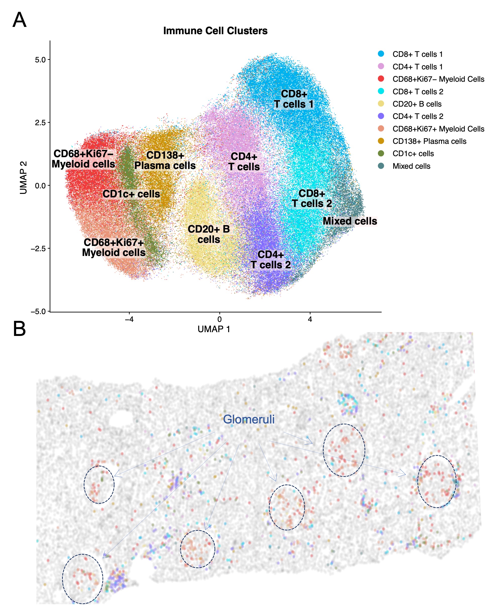
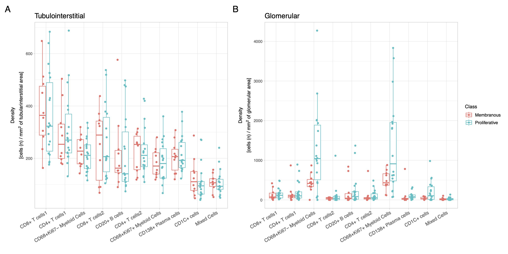
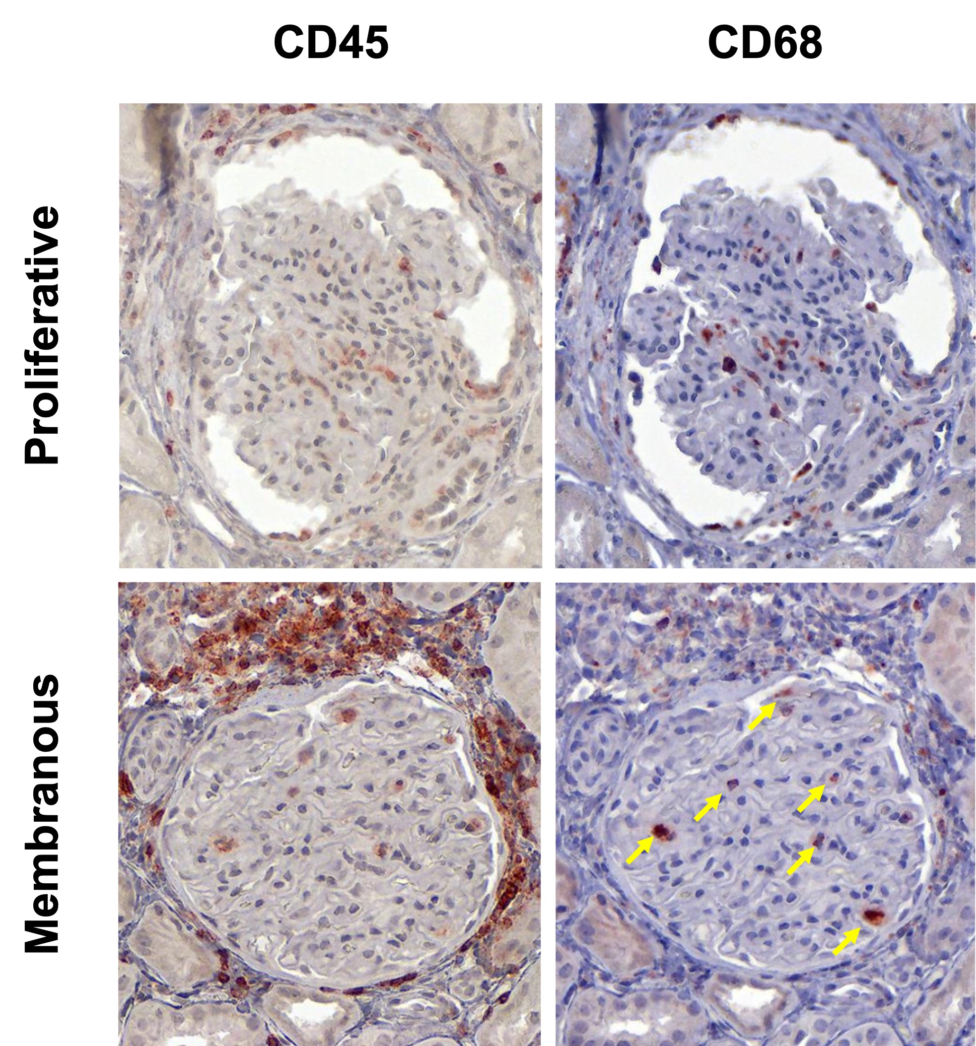
Results: In this initial analysis, we included 29 clinically indicated kidney biopsies classified as LN (13 pure proliferative, 10 pure membranous, 5 mixed, and 1 ISN class II). Patients were 79% female, 34% White, 31% Black, 10% Asian, and 24% identified with Other race/ethnicity. We detected 182,783 CD45+ cells out of 1,913,845 cell objects. Our analysis identified 10 immune cell clusters at low resolution (Figure 1A). Figure 1B displays the tissue distribution of each cell subset. B and T lymphocytes dominated the tubulointerstitium. The CD68+ myeloid subsets were the predominant cell type in the glomeruli (Figure 2). More than half of CD68+ cells expressed Ki67 indicating active proliferation. Surprisingly, we identified intraglomerular CD68+ cells (including endocapillary) also in patients with pure membranous LN, but at a lower tissue density than proliferative LN (Figure 2). Figure 3 demonstrates intraglomerular CD68+ cells in a biopsy classified as pure membranous LN by two experienced renal pathologists.

Conclusion: sIHC can be successfully employed to perform multiplexed whole slide analysis harnessing both the subcellular resolution (brightfield) and the reliability of IHC. Our analysis revealed intraglomerular CD68+ myeloid cells in pure membranous LN. By traditional clinical pathology, intraglomerular / endocapillary immune cells characterize proliferative LN and are not consistent with pure membranous LN. These findings implicate macrophages/monocytes in the glomerular disease in membranous LN with therapeutic implications. The analysis of 90 additional biopsies and a myeloid-focused panel is underway to validate and extend these findings.

Supporting image 1

Figure 1. Phenotype and spatial distribution of intrarenal immune cells. (A) UMAP of CD45+ cells (n=182,783, 29 patients) indicating the low-resolution cluster annotation. (B) Digital reproduction of a representative biopsy displaying the distribution of the cells clusters (colors matching panel A). The biopsy in panel B was classified as ISN class III, NIH Activity Index 6, NIH Chronicity Index 3.

Supporting image 2

Figure 2. Myeloid cells dominate intraglomerular inflammation, including membranous LN. Box plots displaying the density (cells / area) of the immune cell clusters in the tubulointerstitium (A) and glomeruli (B) according to ISN class group. Both CD68+ myeloid cell clusters showed a statistically significant higher intraglomerular density compared to all other clusters in both proliferative and membranous LN (p<0.05, Wilcoxon test). Proliferative LN includes ISN class III and IV +/- V (n=18); membranous LN indicate pure ISN class V (n=10).

Supporting image 3

Figure 3. Intraglomerular CD68+ monocyte/macrophages in proliferative and membranous LN. Immunohistochemistry images displaying the expression of CD45 and CD68 in a representative glomerulus from a patient with proliferative LN (upper panels) and a patient with pure membranous LN (lower panels). The yellow arrows indicate intraglomerular CD68+ cells in membranous LN.


Disclosures: C. Lee: None; C. Marlin: None; X. Yang: None; T. Stephens: None; A. Celia: None; J. Hodgin: AstraZeneca, 5, 6, Eli Lilly, 5, Gilead, 5, Janssen, 5, Moderna, 5, Novo Nordisk, 5, Regeneron, 5; P. Izmirly: None; H. Belmont: Alexion, 6, Aurinia, 6; J. Buyon: Bristol-Myers Squibb(BMS), 2, GlaxoSmithKlein(GSK), 2, Related Sciences, 1; C. Putterman: Equillium, 2, KidneyCure, 1, Progentec, 2; J. James: Bristol-Myers Squibb(BMS), 5, GlaxoSmithKlein(GSK), 2, Novartis, 2, Progentec Biosciences, 5; t. Accelerating Medicines Partnership in RA/SLE: None; M. Petri: Alexion, 1, Amgen, 1, AnaptysBio, 1, Annexon Bio, 1, Argenx, 1, Arhros-Focus Med/Ed, 6, AstraZeneca, 1, 5, Aurinia, 1, 5, 6, Axdev, 1, Biogen, 1, Boxer Capital, 2, Cabaletto Bio, 2, Caribou Biosciences, 2, CVS Health, 1, Eli Lilly, 1, 5, Emergent Biosolutions, 1, Exagen, 5, Exo Therapeutics, 2, Gilead Biosciences, 2, GlaxoSmithKlein(GSK), 1, 5, 6, Horizon Therapeutics, 2, Idorsia Pharmaceuticals, 2, IQVIA, 1, Janssen, 1, 5, Kira Pharmaceuticals, 2, MedShr, 6, Merck/EMD Serono, 1, Momenta Pharmaceuticals, 2, Nexstone Immunology, 2, Nimbus Lakshmi, 2, Proviant, 2, Sanofi, 2, Sinomab Biosciences, 2, Thermofisher, 5, UCB, 2; J. Guthridge: None; A. Rosenberg: None; A. Fava: Annexon Biosciences, 2, Sanofi, 1.

To cite this abstract in AMA style:

Lee C, Marlin C, Yang X, Stephens T, Celia A, Hodgin J, Izmirly P, Belmont H, Buyon J, Putterman C, James J, Accelerating Medicines Partnership in RA/SLE t, Petri M, Guthridge J, Rosenberg A, Fava A. Single-cell Spatial Proteomics Identifies Intraglomerular Myeloid Cells in Membranous Lupus Nephritis [abstract]. Arthritis Rheumatol. 2023; 75 (suppl 9). https://acrabstracts.org/abstract/single-cell-spatial-proteomics-identifies-intraglomerular-myeloid-cells-in-membranous-lupus-nephritis/. Accessed .
  • Tweet
  • Click to email a link to a friend (Opens in new window) Email
  • Click to print (Opens in new window) Print

« Back to ACR Convergence 2023

ACR Meeting Abstracts - https://acrabstracts.org/abstract/single-cell-spatial-proteomics-identifies-intraglomerular-myeloid-cells-in-membranous-lupus-nephritis/

Advanced Search

Your Favorites

You can save and print a list of your favorite abstracts during your browser session by clicking the “Favorite” button at the bottom of any abstract. View your favorites »

All abstracts accepted to ACR Convergence are under media embargo once the ACR has notified presenters of their abstract’s acceptance. They may be presented at other meetings or published as manuscripts after this time but should not be discussed in non-scholarly venues or outlets. The following embargo policies are strictly enforced by the ACR.

Accepted abstracts are made available to the public online in advance of the meeting and are published in a special online supplement of our scientific journal, Arthritis & Rheumatology. Information contained in those abstracts may not be released until the abstracts appear online. In an exception to the media embargo, academic institutions, private organizations, and companies with products whose value may be influenced by information contained in an abstract may issue a press release to coincide with the availability of an ACR abstract on the ACR website. However, the ACR continues to require that information that goes beyond that contained in the abstract (e.g., discussion of the abstract done as part of editorial news coverage) is under media embargo until 10:00 AM ET on November 14, 2024. Journalists with access to embargoed information cannot release articles or editorial news coverage before this time. Editorial news coverage is considered original articles/videos developed by employed journalists to report facts, commentary, and subject matter expert quotes in a narrative form using a variety of sources (e.g., research, announcements, press releases, events, etc.).

Violation of this policy may result in the abstract being withdrawn from the meeting and other measures deemed appropriate. Authors are responsible for notifying colleagues, institutions, communications firms, and all other stakeholders related to the development or promotion of the abstract about this policy. If you have questions about the ACR abstract embargo policy, please contact ACR abstracts staff at [email protected].

Wiley

  • Online Journal
  • Privacy Policy
  • Permissions Policies
  • Cookie Preferences

© Copyright 2025 American College of Rheumatology