Session Information
Session Type: Poster Session B
Session Time: 9:00AM-11:00AM
Background/Purpose: Fibrosis is characterized by collagen deposition, fibro/myofibroblast (MYOFB) accumulation, and extracellular matrix remodeling. In some subtypes of cutaneous lupus erythematosus (CLE), particularly discoid lupus erythematosus (DLE) scarring alopecia and fibrotic lesional resolution may be widespread with no effective treatment. However, mechanisms of scar formation in CLE are not understood. We have shown that epidermal-directed overexpression of murine Vgll3 causes fibrotic-appearing skin lesions suggestive of CLE, and thus, we aimed to explore the role of Vgll3 in cutaneous fibrosis.
Methods: 2–3-month-old male and female transgenic (TG) mice overexpressing Vgll3 in the epidermis under the K5 promoter were compared to wildtype (WT) C57Bl/6 mice (n=3-5 per group). Fibrotic biomarkers of human DLE and scleroderma were compared via immunohistochemistry. In addition, single-cell-RNA-sequencing (scRNA-seq) via 10x platform of lesional and nonlesional skin was completed to investigate the transcriptomes of the potential cellular fibrotic players. Several subclusters of fibroblasts (FBs), MYOFBs, and T cells were identified and fibrotic markers for each FBs subcluster were analyzed in lesional and non-lesional skin.
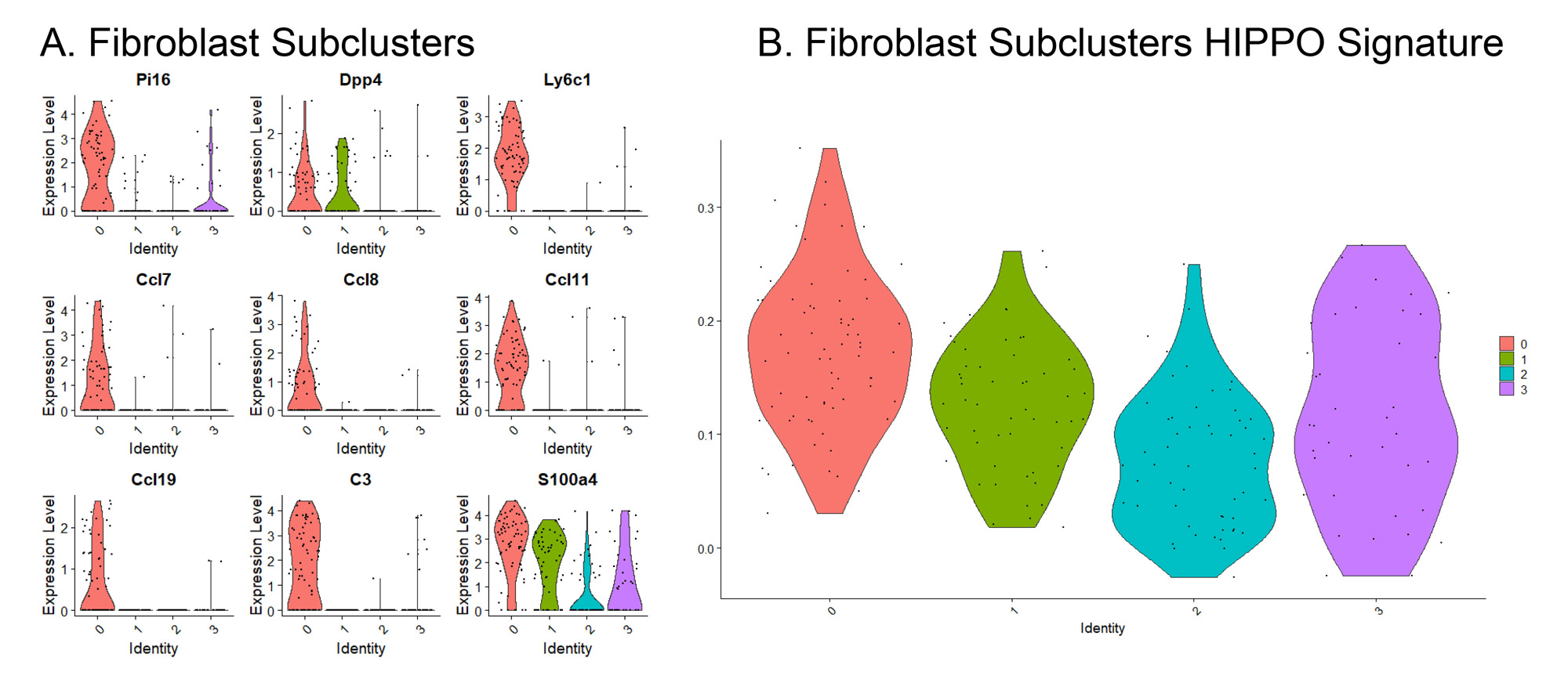
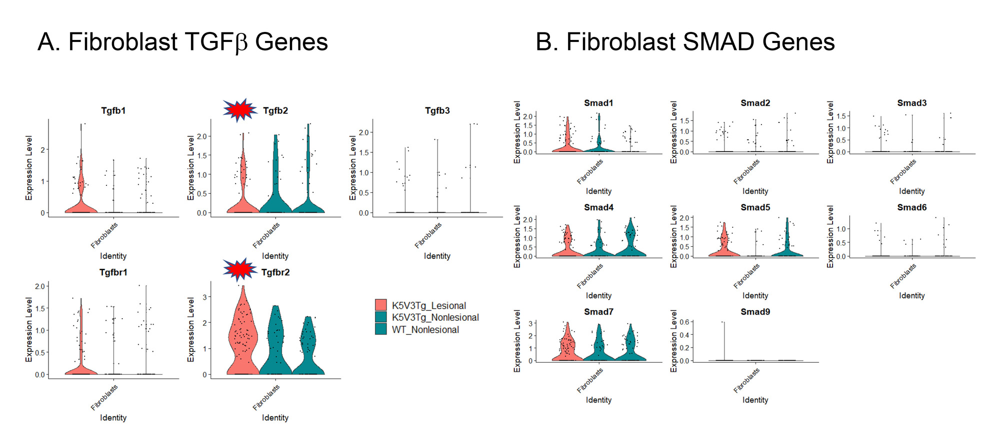
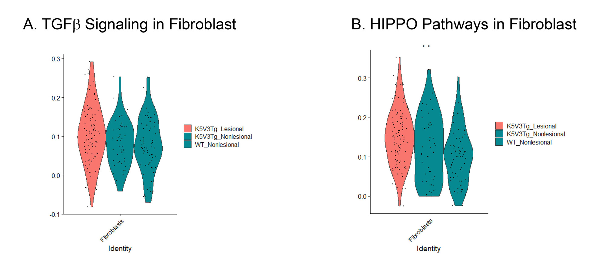
Results: 2–3-month-old male and female transgenic (TG) K5-Vgll3 exhibit cutaneous inflammation and fibrosis as evidenced by trichrome staining. TG lesional skin demonstrated a significant increase in fibrotic markers (Acta2, Col1a1, Col1a2, Tgfb1, Ctgf) and pro-fibrotic cytokines (Il4, Il13) also found in human DLE and scleroderma lesions compared to non-lesional skin. ScRNA-seq of lesional and nonlesional skin from TG vs WT skin showed increased inflammatory infiltrate in lesional TG greater than nonlesional TG, and nonlesional TG, higher than WT skin, which was verified by immunohistochemistry. Increased myeloid cells in lesional TG was higher than nonlesional TG and WT skin. Increased inflammatory gene expression (Nfkb1, Cxcr4, Cxcl2, Il1b, Tnf, Cd14), and CD4+ T cells in lesional TG skin exhibited greater Foxp3, Gata3, and Tgfb1 expression than nonlesional TG and WT skin. Additionally, lesional TG skin exhibited higher expression of Tgfb1,Col1a1, and Col1a2 compared to nonlesional TG and WT mouse skin (transcript and protein). Further, we identified a FB subcluster unique to lesional TG skin representing adventitial Pi16+ FB which expressed chemokines (Ccl7, Ccl8, Ccl11, Ccl19) and exhibited higher MHC class I pathway genes (Fig.1.A) and a higher Hippo signature (Fig.1.B). Finally, increased expression of Hippo and TGFβ pathway-regulated genes in FBs (Figs.2-3), and keratinocytes from lesional and nonlesional TG skin was seen, suggesting Hippo target dysregulation is an early event in fibrosis driven by epidermal Vgll3 that may drive pathologic myofibroblasts differentiation.
Conclusion: Overall, we have linked Vgll3–driven dysregulation of the Hippo and TGFβ pathways to fibrotic phenotypes, which may provide a critical novel target for the alleviation of disfiguring fibrosis in CLE.
A) Lesional Vgll3 TG specific-fibroblasts (FB) subcluster (subcluster 0) express Pi16+ adventitial FB phenotype and express chemokines uniquely. This subcluster Pi16+ adventitial FB (disease FB) is unique to lesional Vgll3 TG skin which express chemokines (CCL7, CCL8, CCL11, CCL19) and exhibit higher MHC class I pathway genes. B) Cluster 0 has highest HIPPO Signature.
B) Lesional Vgll3 TG fibroblast have higher HIPPO pathways score compared to nonlesional.
To cite this abstract in AMA style:
Gharaee-Kermani M, Billi A, Martens J, Hildebrandt M, Victory A, Maz M, Loftus S, Kahlenberg J, Gudjonsson J. Single-Cell RNA Sequencing Reveals Keratinocytes and Fibroblasts Drive Hippo and TGFb Dysregulation and Fibrosis in Epidermal Overexpression of VGLL3 in Cutaneous Lupus [abstract]. Arthritis Rheumatol. 2023; 75 (suppl 9). https://acrabstracts.org/abstract/single-cell-rna-sequencing-reveals-keratinocytes-and-fibroblasts-drive-hippo-and-tgfb-dysregulation-and-fibrosis-in-epidermal-overexpression-of-vgll3-in-cutaneous-lupus/. Accessed .« Back to ACR Convergence 2023
ACR Meeting Abstracts - https://acrabstracts.org/abstract/single-cell-rna-sequencing-reveals-keratinocytes-and-fibroblasts-drive-hippo-and-tgfb-dysregulation-and-fibrosis-in-epidermal-overexpression-of-vgll3-in-cutaneous-lupus/


