ACR Meeting Abstracts

ACR Meeting Abstracts

  • Meetings
    • ACR Convergence 2024
    • ACR Convergence 2023
    • 2023 ACR/ARP PRSYM
    • ACR Convergence 2022
    • ACR Convergence 2021
    • ACR Convergence 2020
    • 2020 ACR/ARP PRSYM
    • 2019 ACR/ARP Annual Meeting
    • 2018-2009 Meetings
    • Download Abstracts
  • Keyword Index
  • Advanced Search
  • Your Favorites
    • Favorites
    • Login
    • View and print all favorites
    • Clear all your favorites
  • ACR Meetings

Abstract Number: 0949

Single-Cell RNA Sequencing of APS Skin Reveals Endothelial Pathology and Cellular Interactions

Hui Shi1, Allison Billi2, Rachael Wasikowski1, Claire Hoy3, Kelsey Gockman1, Lam Tsoi1, Johann Gudjonsson2, Pei-Suen Tsou1 and Jason Knight1, 1University of Michigan, Ann Arbor, MI, 2Department of Dermatology, University of Michigan, Ann Arbor, MI, 3University of Michigan, Ann Arbor

Meeting: ACR Convergence 2021

Keywords: antiphospholipid syndrome, cellular interaction, endothelial cells, livedo reticularis, single cell RNA sequencing

  • Tweet
  • Click to email a link to a friend (Opens in new window) Email
  • Click to print (Opens in new window) Print
Session Information

Date: Sunday, November 7, 2021

Title: Abstracts: Antiphospholipid Syndrome (0948–0951)

Session Type: Abstract Session

Session Time: 9:15AM-9:30AM

Background/Purpose: Although antiphospholipid syndrome (APS) regularly presents with discrete thrombotic events, many patients will also acquire organ damage over time secondary to occlusive neointimal formation in small vessels. The presence of livedo reticularis is predictive of various “internal” manifestations of APS including cerebrovascular disease. Although the interplay between antiphospholipid antibodies (aPL) and endothelial cells is likely a key driver of APS vasculopathy in skin and other organs, APS microvascular endothelial cells have been studied only sparsely. Here, we employed skin biopsy and sc-RNAseq to characterize APS endothelial cells with an eye toward identifying the dysregulated pathways and cellular crosstalk that may conspire to promote APS vasculopathy over time.

Methods: We applied sc-RNAseq to skin biopsies of three primary APS patients with livedo reticularis and four healthy controls with neither APS nor livedo. Uniform manifold approximation and projection (UMAP) was used to discriminate cell types, and differential gene expression analysis was identified. Pathway enrichment was determined by Ingenuity Pathway Analysis. Ligand-receptor pairs from the Database of Interacting Proteins were used to identify potential intra- and inter-cellular communication. Immunohistochemistry (IHC) was performed to verify protein expression in APS and control skin. Control human MVECs were cultured with APS sera in pursuit of discerning transcriptional expression of upregulated genes.

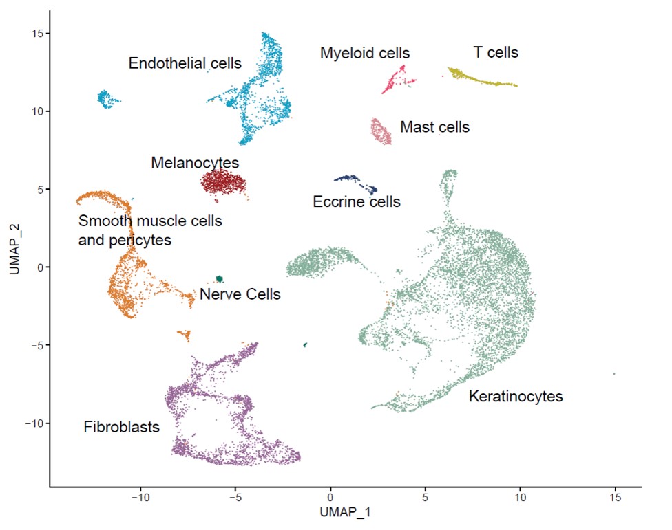
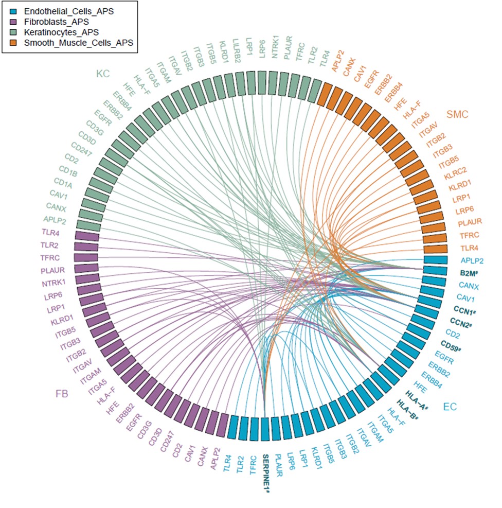
Results: We identified ten cell types in skin (Figure 1). Within the main endothelial cell population, there were four distinct subsets that we named Arterial, Capillary I, Capillary II, and Venous based on differential expression of key genes (Figure 2). We compared gene expression profiles between APS and control endothelial cells and found significant upregulation of various genes associated with cell proliferation, most notably CCN1 (CYR61), CCN2 (CTGF), and SERPINE1 (PAI-1) in APS. These changes were most pronounced in the Capillary I and Venous subsets. By pathway analysis, we found the APS endothelial cell expression profile to be enriched for Hippo-YAP1/TAZ and TGF-β pathways. Anchored by differentially-expressed ligands of APS endothelial cells, interaction analysis suggested strong communication between endothelial cells (especially ligands CCN1 and CCN2) and both pericytes/smooth muscle cells and fibroblasts (Figure 3). IHC staining confirmed higher expression of CCN1 and CCN2 in APS skin as compared with control skin. In these same biopsies, APS endothelial cells were more likely to display YAP1 nuclear translocation as compared with control endothelial cells. In vitro, culture of control MVECs with APS sera upregulated CCN1, CCN2, and SERPINE1.

Conclusion: To our knowledge, this is the first direct analysis of gene expression in APS endothelial cells and local interacting cells. These data suggest that APS endothelial cells demonstrate a pattern of ligand gene expression with potential to drive the proliferation of neighboring cells including pericytes/smooth muscle cells and fibroblasts, which may have significant relevance for the neointimal formation of APS vasculopathy.

Cell type-specific subsets in APS and control skin.

Endothelial cell subsets in APS and control skin.

Interactions between ligands upregulated in APS endothelial cells (bolded) and receptors expressed by endothelial cells and other cell types. EC=endothelial cells, SMC=pericytes/smooth muscle cells, KC=keratinocytes, and FB=fibroblasts.


Disclosures: H. Shi, None; A. Billi, None; R. Wasikowski, None; C. Hoy, None; K. Gockman, None; L. Tsoi, None; J. Gudjonsson, Almirall, 5, Eli Lilly, 5, BMS, 5; P. Tsou, None; J. Knight, None.

To cite this abstract in AMA style:

Shi H, Billi A, Wasikowski R, Hoy C, Gockman K, Tsoi L, Gudjonsson J, Tsou P, Knight J. Single-Cell RNA Sequencing of APS Skin Reveals Endothelial Pathology and Cellular Interactions [abstract]. Arthritis Rheumatol. 2021; 73 (suppl 9). https://acrabstracts.org/abstract/single-cell-rna-sequencing-of-aps-skin-reveals-endothelial-pathology-and-cellular-interactions/. Accessed .
  • Tweet
  • Click to email a link to a friend (Opens in new window) Email
  • Click to print (Opens in new window) Print

« Back to ACR Convergence 2021

ACR Meeting Abstracts - https://acrabstracts.org/abstract/single-cell-rna-sequencing-of-aps-skin-reveals-endothelial-pathology-and-cellular-interactions/

Advanced Search

Your Favorites

You can save and print a list of your favorite abstracts during your browser session by clicking the “Favorite” button at the bottom of any abstract. View your favorites »

All abstracts accepted to ACR Convergence are under media embargo once the ACR has notified presenters of their abstract’s acceptance. They may be presented at other meetings or published as manuscripts after this time but should not be discussed in non-scholarly venues or outlets. The following embargo policies are strictly enforced by the ACR.

Accepted abstracts are made available to the public online in advance of the meeting and are published in a special online supplement of our scientific journal, Arthritis & Rheumatology. Information contained in those abstracts may not be released until the abstracts appear online. In an exception to the media embargo, academic institutions, private organizations, and companies with products whose value may be influenced by information contained in an abstract may issue a press release to coincide with the availability of an ACR abstract on the ACR website. However, the ACR continues to require that information that goes beyond that contained in the abstract (e.g., discussion of the abstract done as part of editorial news coverage) is under media embargo until 10:00 AM ET on November 14, 2024. Journalists with access to embargoed information cannot release articles or editorial news coverage before this time. Editorial news coverage is considered original articles/videos developed by employed journalists to report facts, commentary, and subject matter expert quotes in a narrative form using a variety of sources (e.g., research, announcements, press releases, events, etc.).

Violation of this policy may result in the abstract being withdrawn from the meeting and other measures deemed appropriate. Authors are responsible for notifying colleagues, institutions, communications firms, and all other stakeholders related to the development or promotion of the abstract about this policy. If you have questions about the ACR abstract embargo policy, please contact ACR abstracts staff at [email protected].

Wiley

  • Online Journal
  • Privacy Policy
  • Permissions Policies
  • Cookie Preferences

© Copyright 2025 American College of Rheumatology