ACR Meeting Abstracts

ACR Meeting Abstracts

  • Meetings
    • ACR Convergence 2024
    • ACR Convergence 2023
    • 2023 ACR/ARP PRSYM
    • ACR Convergence 2022
    • ACR Convergence 2021
    • ACR Convergence 2020
    • 2020 ACR/ARP PRSYM
    • 2019 ACR/ARP Annual Meeting
    • 2018-2009 Meetings
    • Download Abstracts
  • Keyword Index
  • Advanced Search
  • Your Favorites
    • Favorites
    • Login
    • View and print all favorites
    • Clear all your favorites
  • ACR Meetings

Abstract Number: 1703

Single Cell and Spatial Transcriptomics Implicate Vascular Cell Orchestration of Inflammatory Changes After Ultraviolet Light Exposure

Rachael Wasikowski1, Erin Theisen2, Jie An3, Lam C. Tsoi1, Ksenia Anufrieva4, Kevin Wei5, Johann Gudjonsson6 and Keith Elkon3, 1Michigan, Dept. of Dermatology, Ann Arbor, MI, 2Brigham and Women’s Hospital and Harvard Medical School, Boston, MA, 3University of Washington, Seattle, WA, 4BWH, Cambridge, MA, 5Brigham and Women's Hospital at Harvard Medical School, Boston, MA, 6University of Michigan, Ann Arbor, MI

Meeting: ACR Convergence 2024

Keywords: chemokines, complement, Inflammation, skin, Systemic lupus erythematosus (SLE)

  • Tweet
  • Email
  • Print
Session Information

Date: Sunday, November 17, 2024

Title: Abstracts: Innate Immunity: Molecular Insights Into Immune Dysregulation

Session Type: Abstract Session

Session Time: 3:00PM-4:30PM

Background/Purpose: Skin sensitivity to sunlight affects ~70% of systemic lupus erythematosus (SLE). In addition, ultraviolet radiation (UVR) can stimulate systemic disease flares, but how this occurs is unknown. To systematically examine the acute effects of UVR, we performed single cell nuclear RNA sequencing (snRNAseq) and a spatial and kinetic analysis of gene expression in the skin following UVR.

Methods: Shaved mouse skin received a single exposure of 500 mJ/cm2 UVB, and biopsies were taken at times 0 and 12 hrs (snRNAseq) and then 0, 6,  and 24 hours after UVB exposure (n=3 per group) for spatial transcriptome analysis. snRNAseq employed Illumina NextSeq protocols with bioinformatics utilizing Monocle and Seurat pipelines. For spatial analysis, fixed tissue was stained with antibodies to CD45 and to cytokeratin (CK) to mark immune cells and keratinocytes, respectively and processed on the GeoMx digital spatial profiler.  Mouse whole transcriptome probe set was used for next-generation sequence analysis and processed on the nanoString Software Suite 2.5.1.145. Pathway analysis was determined by Ingenuity (IPA).

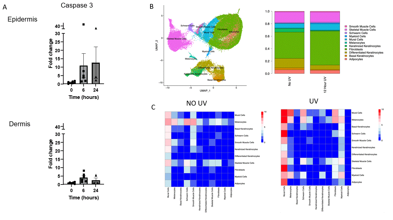
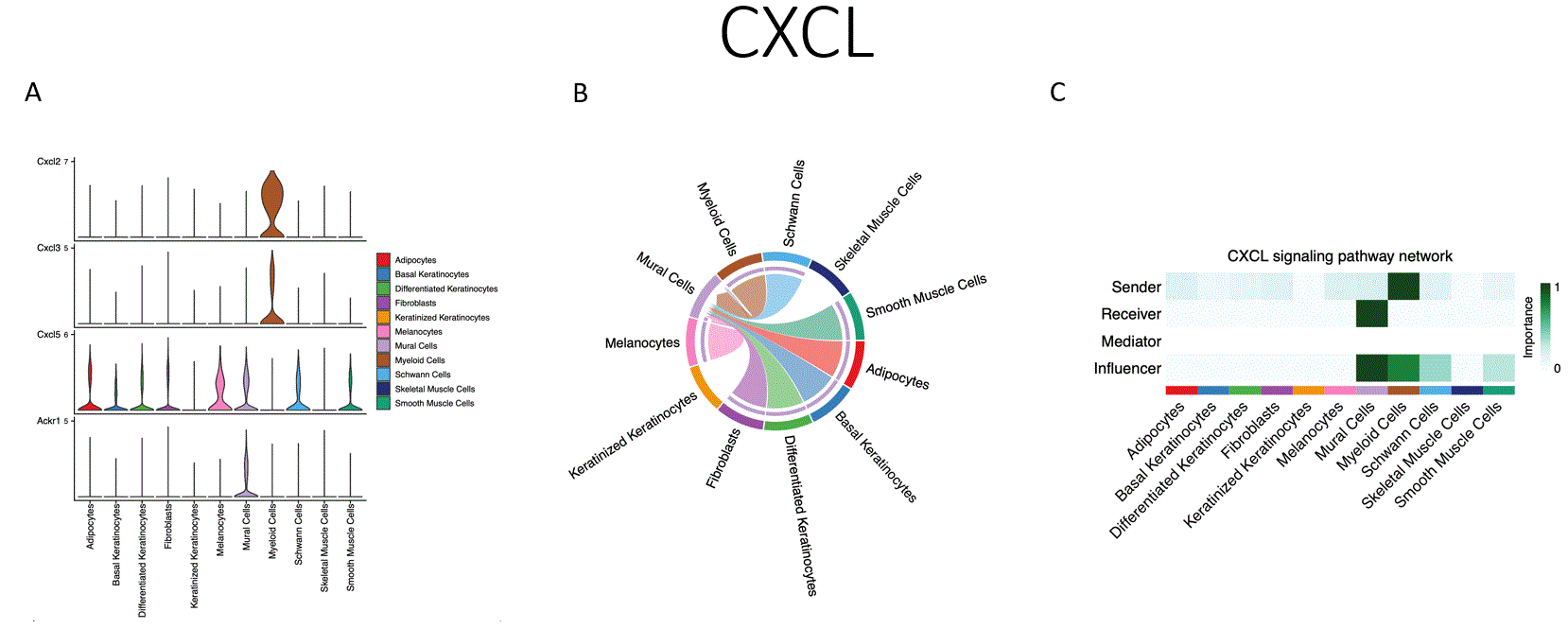
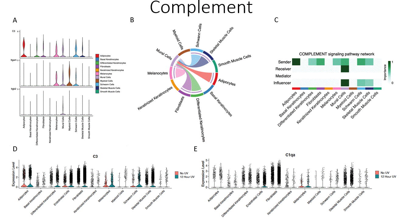
Results: UVR was accompanied by increased cellular apoptosis (caspase 3 staining), which peaked at 6 hours (Fig.1). SnRNAseq with 51,162 cells identified 12 cellular subtypes and revealed a shift toward increased myeloid and endothelial cell numbers, with decreased proportions of differentiated and keratinized keratinocytes. The most marked changes in gene expression were observed in melanocytes (MLNs), followed by endothelial cells (ECs) and keratinocytes, with lesser shifts in fibroblast and myeloid cells.  Enriched biological processes shared across multiple cell types include enriched cytokine signaling in the immune system, neutrophil degranulation, senescence-associated secretory phenotype, and inflammasome pathway activation.  Receptor ligand interactions demonstrated shifts towards increased activity of ECs, MLNs, and myeloid cells.  Unique pathways increased with UVB exposure included CXCL chemokines (CXCL1, CXCL2, and CXCL5, Fig.2), increased CCL chemokine expression (CCL2, CCL6, CCL7, and CCL9), and complement components (C3, C4, and C1qa, Fig. 3).  The major contributors to chemokine gene expression were myeloid and ECs, whereas C3 gene expression was seen across multiple cell types.  These data were validated by spatial profiling (GeoMX) of the epidermis, dermis, and subcutaneous tissue.  The most marked changes were in the epidermal and superficial dermal compartment with IPA highlighting apoptosis, UV-response, unfolded protein response, and p53 pathway activation and also DEGs involving leukocyte activation, cellular defense response, and regulation of reactive oxygen species.

Conclusion: Following UVR, cell stress leads to epithelial cell death and activation of inflammatory pathways. Interestingly, ECs appear to be central in orchestrating UVR inflammatory responses (Figs. 1,2,3C) that include stimulation of chemokines and complement, which in turn facilitate myeloid infiltration and immune activation. Many of these genes and pathways have been shown to be abnormal in diseases such as lupus. 

Supporting image 1

Fig. 1. Apoptosis and single-cell analyses from UVR exposed mouse skin. Change in Caspase_3 staining in UVR mouse skin at different locations and time points (A). UMAP of single-cell data obtained from UVR-exposed skin at 12 hours and cell proportion in UVR-treated vs. unexposed skin (B). Heatmap of immune-related receptor-ligand interactions in unexposed vs. UVR-treated skin at 12 hours. The colors represent the total number of receptor-ligand pairs (C).

Supporting image 2

Fig. 2 Activated CXCL family chemokines following UVR at 12 hours. Violin plots of Cxcl2, Cxcl3, Xcxl5, and ACKR1 in UVR exposed skin (A). Circos plot of interactions related to CXCL pathway signaling between endothelial cells (mural cells) and other cell types in UVR-exposed skin (B). CXCL signaling pathway network in UVR-exposed skin (C).

Supporting image 3

Fig. 3. Activated Complement family following UVR at 12 hours. Violin plots of C3, Itgam, and Itgb2 in UVR exposed skin (A). Circos plot of interactions related to complement pathway signaling between endothelial cells (mural cells) and other cell types in UVR-exposed skin (B). Complement signaling pathway network in UVR-exposed skin (C). The expression of C3 (D) and C1qa (D), as violin plots of UVR-exposed skin (green) vs control non-exposed skin (red) at 12 hours.


Disclosures: R. Wasikowski: None; E. Theisen: None; J. An: None; L. Tsoi: Janssen, 5; K. Anufrieva: None; K. Wei: 10X Genomics, 5, Gilead sciences, 2, Merck/MSD, 5, Mestag, 2, santa ana bio, 2; J. Gudjonsson: AbbVie/Abbott, 12, support, Boehringer-Ingelheim, 12, support, Bristol-Myers Squibb(BMS), 12, support, Eli Lilly, 12, support, Janssen, 12, support, Novartis, 12, support; K. Elkon: None.

To cite this abstract in AMA style:

Wasikowski R, Theisen E, An J, Tsoi L, Anufrieva K, Wei K, Gudjonsson J, Elkon K. Single Cell and Spatial Transcriptomics Implicate Vascular Cell Orchestration of Inflammatory Changes After Ultraviolet Light Exposure [abstract]. Arthritis Rheumatol. 2024; 76 (suppl 9). https://acrabstracts.org/abstract/single-cell-and-spatial-transcriptomics-implicate-vascular-cell-orchestration-of-inflammatory-changes-after-ultraviolet-light-exposure/. Accessed .
  • Tweet
  • Email
  • Print

« Back to ACR Convergence 2024

ACR Meeting Abstracts - https://acrabstracts.org/abstract/single-cell-and-spatial-transcriptomics-implicate-vascular-cell-orchestration-of-inflammatory-changes-after-ultraviolet-light-exposure/

Advanced Search

Your Favorites

You can save and print a list of your favorite abstracts during your browser session by clicking the “Favorite” button at the bottom of any abstract. View your favorites »

All abstracts accepted to ACR Convergence are under media embargo once the ACR has notified presenters of their abstract’s acceptance. They may be presented at other meetings or published as manuscripts after this time but should not be discussed in non-scholarly venues or outlets. The following embargo policies are strictly enforced by the ACR.

Accepted abstracts are made available to the public online in advance of the meeting and are published in a special online supplement of our scientific journal, Arthritis & Rheumatology. Information contained in those abstracts may not be released until the abstracts appear online. In an exception to the media embargo, academic institutions, private organizations, and companies with products whose value may be influenced by information contained in an abstract may issue a press release to coincide with the availability of an ACR abstract on the ACR website. However, the ACR continues to require that information that goes beyond that contained in the abstract (e.g., discussion of the abstract done as part of editorial news coverage) is under media embargo until 10:00 AM ET on November 14, 2024. Journalists with access to embargoed information cannot release articles or editorial news coverage before this time. Editorial news coverage is considered original articles/videos developed by employed journalists to report facts, commentary, and subject matter expert quotes in a narrative form using a variety of sources (e.g., research, announcements, press releases, events, etc.).

Violation of this policy may result in the abstract being withdrawn from the meeting and other measures deemed appropriate. Authors are responsible for notifying colleagues, institutions, communications firms, and all other stakeholders related to the development or promotion of the abstract about this policy. If you have questions about the ACR abstract embargo policy, please contact ACR abstracts staff at [email protected].

Wiley

  • Online Journal
  • Privacy Policy
  • Permissions Policies
  • Cookie Preferences

© Copyright 2025 American College of Rheumatology