
Session Information
Date: Sunday, November 17, 2024
Title: SpA Including PsA – Diagnosis, Manifestations, & Outcomes Poster II
Session Type: Poster Session B
Session Time: 10:30AM-12:30PM
Background/Purpose: Spinal degenerative lesions (DLs) are commonly seen/reported in radiographs and MRI in axSpA. However, the relationship between the different DLs over time has not been established. Our aim was to investigate the relationship between specific DLs over 10 years (10Y) in axSpA.
Methods: Whole spine MRI and cervical and lumbar spine radiographs at baseline, 5Y and 10Y of patients diagnosed with axSpA in the DESIR cohort were assessed for DLs by three central readers blinded to timepoint and to clinical, laboratory or any other imaging information1. Only patients with ≥2 time points available were analyzed. We used multilevel generalised estimating equation (GEE) time-lagged and autoregressive models adjusted for age, sex, HLA-B27 status, BMI, smoking and job type at baseline and biologic therapy during follow-up (time-varying variable). Time-lagged models test the effect of one DL at one time point on another DL in the subsequent time point. Autoregressive means that the models were adjusted for the outcome in the previous time point, which is similar to modelling a change in the outcome. Adjusted odds ratios (aOR) and 95% confidence intervals (95%CI) were reported.
Results: DLs were available for 291 patients on MRI and 323 patients on radiographs (mean age [SD] 34.5 [8.6] years; 47% men). On MRI, significant associations were found between disc degeneration and herniation (aOR=4.0; 95%CI: 3.5-4.7), high intensity zone (1.8; 1.5-2.1) and Modic type 1 lesion (4.7; 3.7-6.1) in the subsequent time point at the same vertebral unit level, after controlling for such a lesion at the previous time point (Table 1). There was also a significant association between Modic type 1 and subsequent Modic type 2 or 3 lesions (54.0; 38.0-76.9). Herniations tended to persist over time (187; 156-224). On radiographs, disc height loss was significantly associated with a subsequent osteophyte (4.3; 3.6-5.1) (Table 2). After adjustments, there was a significant association between having involvement at ≥1 vertebral level and having involvement at multiple vertebral levels or adjacent levels in the subsequent time point on radiographs and on MRI (Figure 1). The presence of a DL on MRI seemed to predict its appearance on subsequent radiographs (2.5; 2.2-2.8).
Conclusion: In this first study providing insight into the long-term relationship of spinal DLs in an inception cohort of axSpA, different DLs are associated with the development of other DLs over time. These findings could help rheumatologists to better understand DLs and their evolution over time.
Reference: 1. de Bruin F et al. RMD Open. 2018;4(1):e000657.
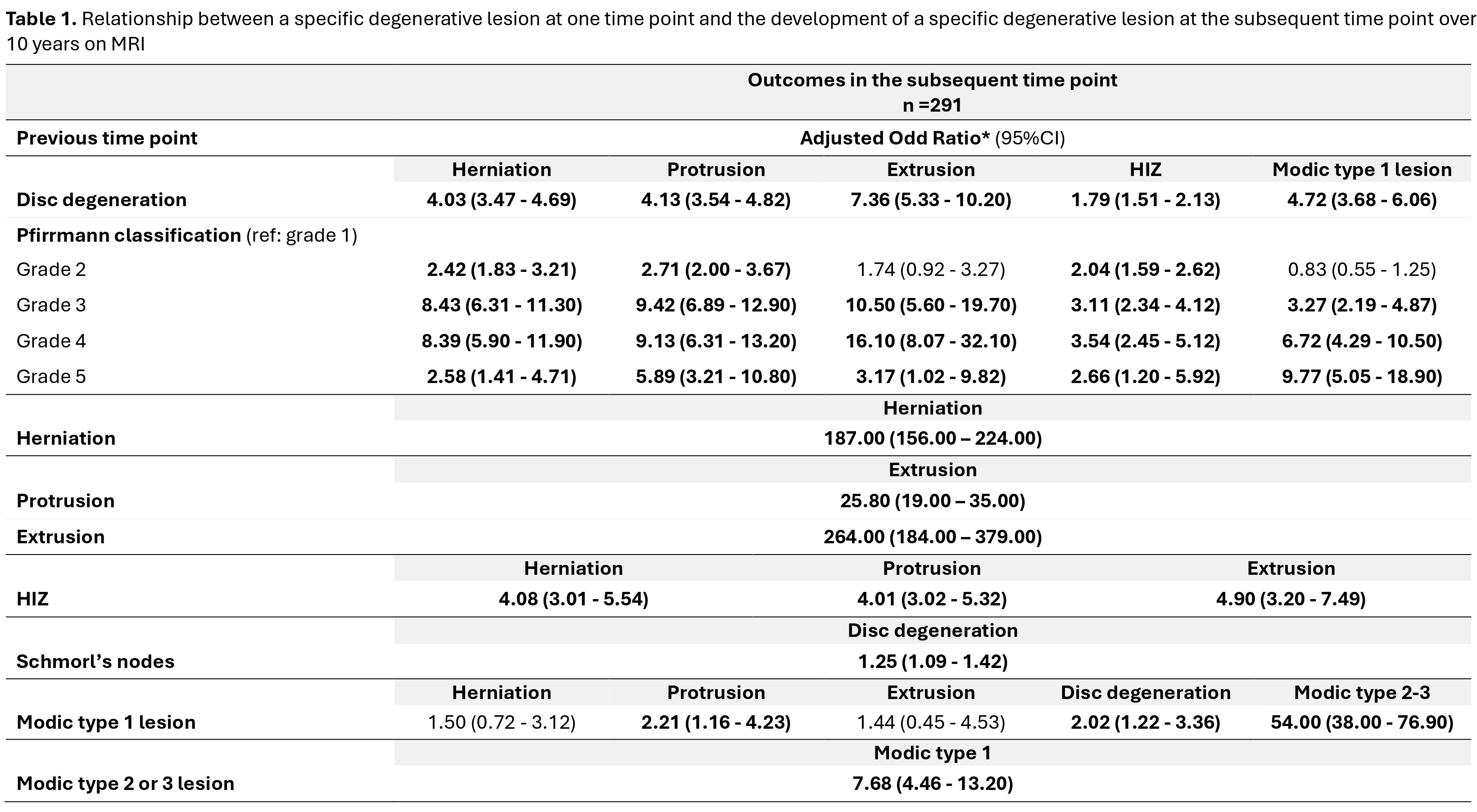
Disc degeneration was scored on a five-point scale (Pfirrmann classification), combining signal loss and loss of height of the intervertebral discs on STIR images, and was defined as a disc with a Pfirrmann classification >2. The high-intensity zone, indicating an annular tear or fissure, is seen as an area of high signal intensity located in the posterior annulus fibrosis on STIR images. We considered bulging of a disc/herniation as either protrusion or extrusion; the distinction is based on whether the maximal diameter in any direction is at the base of the displacement (protrusion) or not (extrusion). We used the Modic classification to assess degenerative lesions of the vertebral endplates. This is a three-point scale with type I defined as bone marrow oedema, type II is described as fatty changes and type III as sclerotic changes. Schmorl’s nodes were defined as an indentation of the (cranial or caudal) endplate with herniation of intervertebral disc material into the vertebra, with or without oedema.
Bold characters indicate significant values. 95%CI: 95% confidence interval; HIZ: high intensity zone.
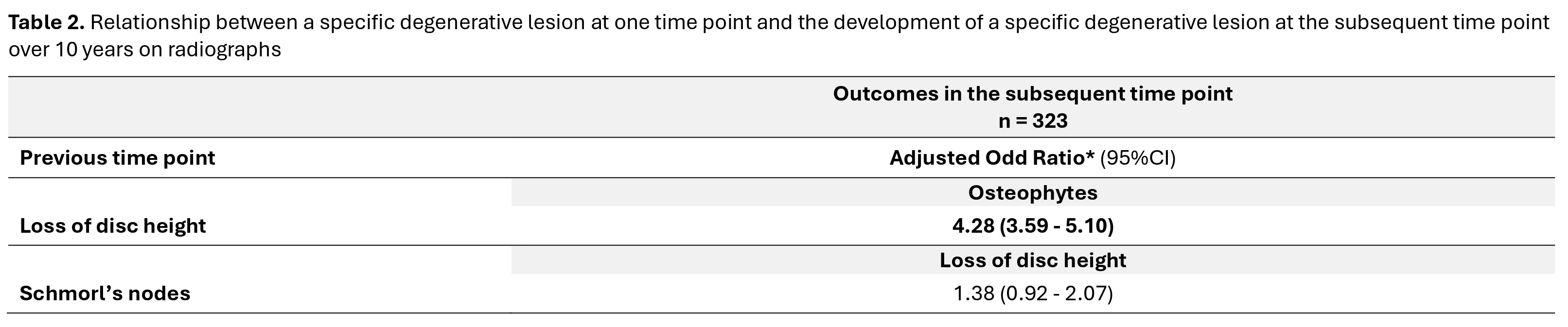
Loss of disc height was defined as narrowing of the disc space in comparison with two adjacent (healthy) discs. Osteophytes were described by reactive bone hypertrophy, seen as bony spurs arising from the vertebral body close to the vertebral endplate in a horizontal configuration (maximum of 45-degree angle with the endplate). Schmorl’s nodes were defined as a radiolucent contour defect of the vertebral endplate with sclerotic margins.
Bold characters indicate significant values. 95%CI: 95% confidence interval.
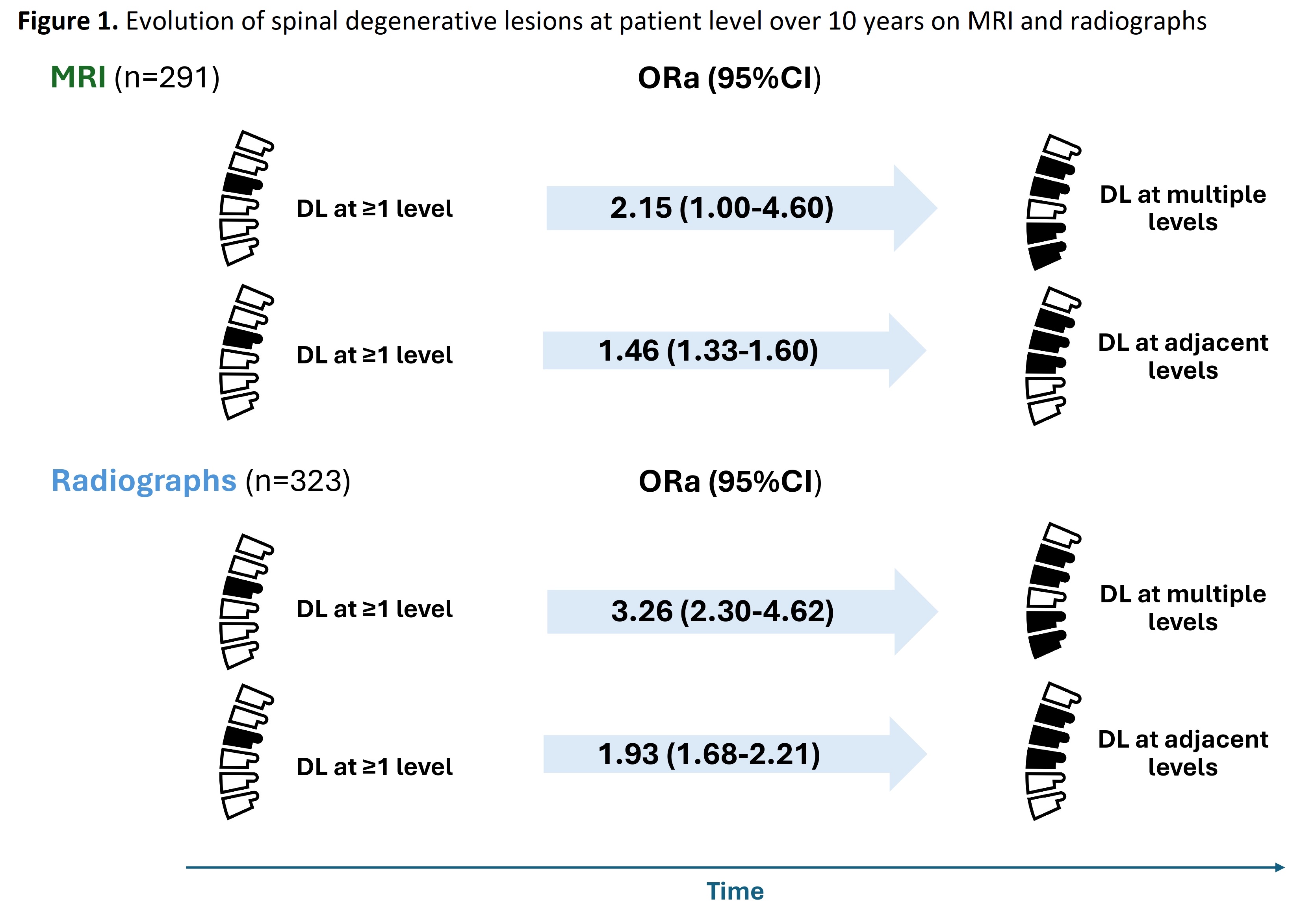
DL: degenerative lesion; ORa: adjusted odd ratio; 95%CI: 95% confidence interval.
To cite this abstract in AMA style:
Pina Vegas L, van Gaalen F, van Lunteren M, Loeuille D, Morizot C, Newsum E, Claudepierre P, SARAUX A, Feydy A, Reijnierse M, van der Heijde D, Ramiro S. Relationship Between Degenerative Lesions over Time in Axial Spondyloarthritis: 10-year Follow-up of the DESIR Cohort [abstract]. Arthritis Rheumatol. 2024; 76 (suppl 9). https://acrabstracts.org/abstract/relationship-between-degenerative-lesions-over-time-in-axial-spondyloarthritis-10-year-follow-up-of-the-desir-cohort/. Accessed .« Back to ACR Convergence 2024
ACR Meeting Abstracts - https://acrabstracts.org/abstract/relationship-between-degenerative-lesions-over-time-in-axial-spondyloarthritis-10-year-follow-up-of-the-desir-cohort/