Session Information
Date: Monday, November 9, 2015
Title: Imaging of Rheumatic Diseases Poster II: X-ray, MRI, PET and CT
Session Type: ACR Poster Session B
Session Time: 9:00AM-11:00AM
Background/Purpose:
Standard assessment of Rheumatoid Arthritis (RA) on MRI relies on semi-quantitative joint space narrowing scores that are not sensitive to cartilage focal lesions or early biochemical damage1,2. MRI T1ρ has been shown to quantitatively evaluate cartilage proteoglycan loss, collagen matrix changes, and water content3. The purpose of our study was to evaluate the feasibility of radiocarpal cartilage T1ρ as a biomarker in RA clinical trials.
Methods:
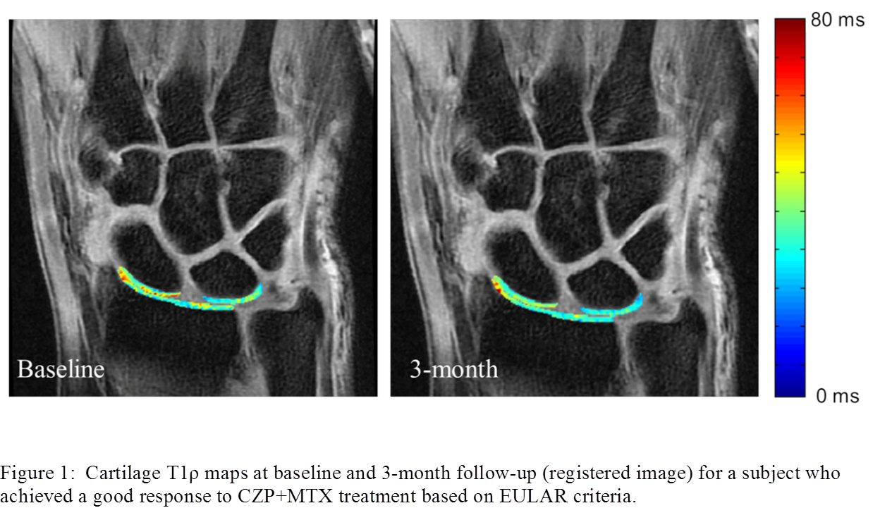
Nine patients were included in this study: 6 RA patients with active disease despite methotrexate (MTX) treatment started on certolizumab pegol (CZP) and 3 RA patients with low disease on MTX treatment alone. 3T MR was performed at baseline (immediately before CZP initiation) and after 3 months for all subjects. A coronal T1ρ sequence was used (Time of Spin-Lock = 0/10/20/50ms; spin lock frequency = 500Hz, resolution 0.21×0.21x3mm) for lunate, scaphoid, and radius cartilage segmentations. All echoes and follow-up images were registered to baseline images with piecewise rigid registration using individual bone masks (Fig. 1). Paired t-tests were used to compare T1ρ values at baseline and 3-month. Progression of T1ρ values (ΔT1ρ) were correlated (Spearman’s rho) with progression of Disease Activity Scores, Michigan Hand Questionnaires, and Health Assessment Questionnaires.
Results:
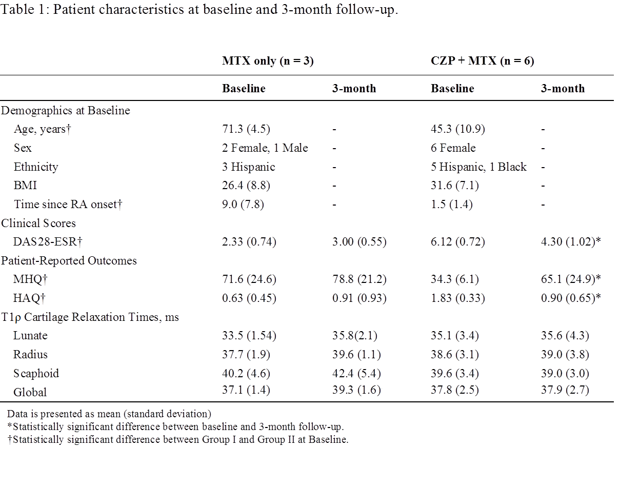
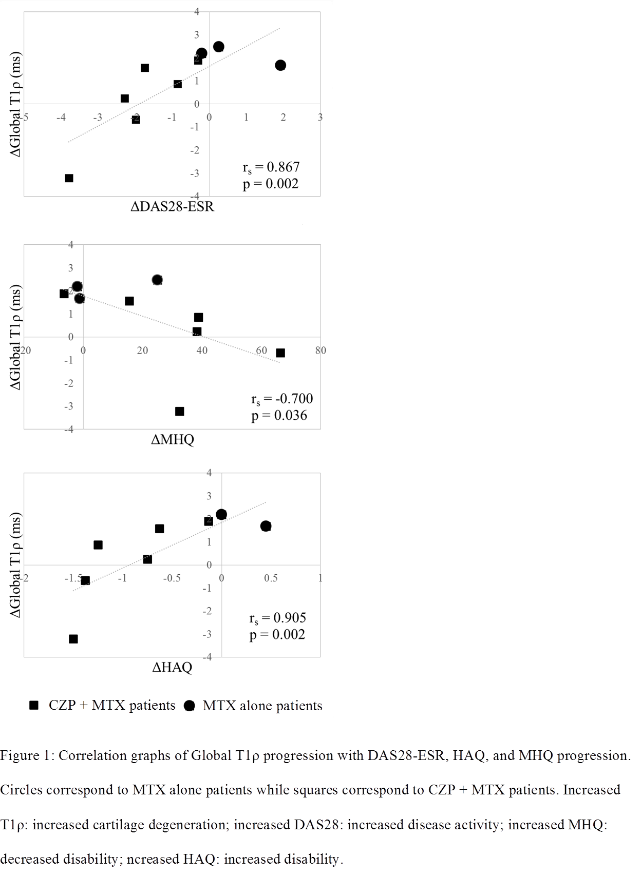
Patient characteristics are presented in Tab. 1. MTX alone patients showed an increase in Global T1ρ (37.4±1.4 to 39.3±1.6 ms) after 3 months while CZP + MTX patients did not show any significant changes in T1ρ. A statistically significant correlation was found between ΔGlobal T1ρ and ΔDAS28-ESR, ΔMHQ, and ΔHAQ (Fig. 2).
Conclusion:
We previously reported excellent in vivo reproducibility of wrist cartilage T1ρ (1.42% – 5.62% CV) 4. The overall non-significant change of cartilage T1ρ at 3 months for CZP + MTX patients suggests cartilage responds positively to anti-TNF treatment through joint structure protection. Despite the small sample size, there are already strong correlations between progression of cartilage T1ρ with DAS28, MHQ, and HAQ, encouraging further research with more patients and time points.
References:
[1] Dohn et al., J of Rheumatol 2014 [2] Duvvuri et al., Magn Reson in Med 1997 [3] Li et al., Osteoarthritis Cartilage 2007 [4] Pedoia et al., ACR Abstract 2014
To cite this abstract in AMA style:
Ku E, Pedoia V, Tanaka M, Heilmeier U, Imboden J, Graf JD, Link TM, Li X. Radiocarpal Cartilage Matrix Changes 3-Months after Anti-TNF Treatment for Rheumatoid Arthritis – Feasibility Study Using MR T1ρ� Imaging [abstract]. Arthritis Rheumatol. 2015; 67 (suppl 10). https://acrabstracts.org/abstract/radiocarpal-cartilage-matrix-changes-3-months-after-anti-tnf-treatment-for-rheumatoid-arthritis-feasibility-study-using-mr-t1%ef%bf%bd-imaging/. Accessed .« Back to 2015 ACR/ARHP Annual Meeting
ACR Meeting Abstracts - https://acrabstracts.org/abstract/radiocarpal-cartilage-matrix-changes-3-months-after-anti-tnf-treatment-for-rheumatoid-arthritis-feasibility-study-using-mr-t1%ef%bf%bd-imaging/


