ACR Meeting Abstracts

ACR Meeting Abstracts

  • Meetings
    • ACR Convergence 2024
    • ACR Convergence 2023
    • 2023 ACR/ARP PRSYM
    • ACR Convergence 2022
    • ACR Convergence 2021
    • ACR Convergence 2020
    • 2020 ACR/ARP PRSYM
    • 2019 ACR/ARP Annual Meeting
    • 2018-2009 Meetings
    • Download Abstracts
  • Keyword Index
  • Advanced Search
  • Your Favorites
    • Favorites
    • Login
    • View and print all favorites
    • Clear all your favorites
  • ACR Meetings

Abstract Number: 2064

Quantifying Cutaneous Dermatomyositis: A Novel 3D Image-based Approach

Nantakarn Pongtarakulpanit1, Tanya Chandra1, shiri keret2, Eugenia Gkiaouraki3, Vladimir Liarski1, Dana Ascherman3, Siamak Mogahadam1, Chester Oddis1 and Rohit Aggarwal4, 1Division of Rheumatology and Clinical Immunology, University of Pittsburgh, Pittsburgh, PA, 2Rheumatology unit, Bnai-Zion medical center and the faculty of Medicine, Technion - Israel Institute of Technology, Haifa, Israel., Atlit, Israel, 3Division of Rheumatology and Clinical Immunology, University of Pittsburgh Medical Center, Pittsburgh, PA, 4Division of Rheumatology and Clinical Immunology, University of Pittsburgh, Pittsburgh, PA, USA, Pittsburgh, PA

Meeting: ACR Convergence 2024

Keywords: dermatomyositis, Imaging, Outcome measures

  • Tweet
  • Click to email a link to a friend (Opens in new window) Email
  • Click to print (Opens in new window) Print
Session Information

Date: Monday, November 18, 2024

Title: Muscle Biology, Myositis & Myopathies – Basic & Clinical Science Poster III

Session Type: Poster Session C

Session Time: 10:30AM-12:30PM

Background/Purpose: Rapid advancement in image analysis now has a critical role in the diagnosis and assessment of skin lesions. The current standard of visual examination and scoring has considerable subjectivity, recall bias for assessing change, and inter-rater reliability, particularly when reported by non-dermatologists. This study assesses the feasibility of 3D image-based assessment to evaluate cutaneous disease activity in dermatomyositis (DM).

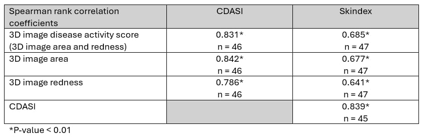
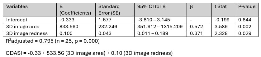
Methods: DM patients seen in the University of Pittsburgh Myositis Center between August 2022 and April 2024 had 3D images of their rashes generated using a 3D camera. Each set of images included 7 “standard” areas (dorsum of left hand, dorsum of right hand, left side of the upper chest, right side of the upper chest, left side of the upper back, right side of the upper back, and face), and “other” involved areas based on rash distribution. DM rashes were manually mapped and measured for area and redness using the 3D image application. A “3D image disease activity score” was calculated based on the percentage of the rashes relative to the total body surface area, multiplied by the degree of redness (based on average red/green coordinate in the CIELAB color Space system). The construct validity and responsiveness of the score were evaluated using the Spearman rank correlation coefficient against a standard in-clinic myositis expert evaluation of “Cutaneous Dermatomyositis Disease Area and Severity Index (CDASI)” and a standard patient-reported outcome termed the “Skindex”. A linear regression model assessed the relationship between the 3D image-derived rash area and redness with the CDASI score.

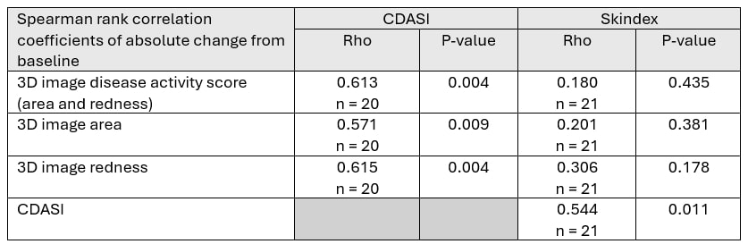
Results: 27 DM patients with 1-2 clinic visits spaced at least 3 months apart were enrolled. Subjects were predominantly female (81.5%) and Caucasian (96.3%). The median (IQR) CDASI and Skindex scores were 6.0 (0.0 – 17.0) and 28.0 (4.0 – 45.0), respectively. For the construct validity, the 3D image disease activity score correlated strongly with the physician-derived CDASI and patient-reported Skindex scores (Spearman rank correlation coefficients 0.83 and 0.67, respectively, p< 0.01). The linear regression analysis identified rash area and redness from 3D images as significant predictors of the CDASI score. The model explained 79.5% of the variance in CDASI scoring (R2 = 0.795). Regarding responsiveness, absolute changes from baseline in 3D image disease activity score correlated strongly with absolute changes in the CDASI score (the Spearman rank correlation coefficient 0.61, P = 0.004).

Conclusion: Our results demonstrate favorable validity and responsiveness in the 3D images for evaluating rash in DM patients. The 3D image-derived rash area and redness are significant predictors of CDASI scores, supporting the utility of 3D imaging in objective disease activity assessments of cutaneous DM.

Supporting image 1

Table 1. The correlations of 3D image parameters with Cutaneous Dermatomyositis Disease Area and Severity Index (CDASI) and Skindex scores

Supporting image 2

Table 2. The correlations of absolute change from the baseline of 3D image parameters with absolute change from the baseline of Cutaneous Dermatomyositis Disease Area and Severity Index (CDASI) and Skindex scores

Supporting image 3

Table 3. A linear regression model with regression coefficients for the relationship between the 3D image-derived rash area and redness with the Cutaneous Dermatomyositis Disease Area and Severity Index (CDASI).


Disclosures: N. Pongtarakulpanit: None; T. Chandra: None; s. keret: None; E. Gkiaouraki: None; V. Liarski: None; D. Ascherman: None; S. Mogahadam: None; C. Oddis: Abcuro, 5, Alexion, 5, Argenx, 5, Boehringer Ingelheim, 5, CSL Behring, 5, EMD Serono, 5, Janssen, 5, Pfizer, 5, Priovant, 5; R. Aggarwal: Alexion, 2, ANI Pharmaceuticals, 2, Argenx, 2, AstraZeneca, 2, Boehringer-Ingelheim, 2, 5, Bristol-Myers Squibb(BMS), 2, 5, CabalettaBio, 2, Capella Bioscience, 2, Capstanx, 2, Corbus, 2, CSL Behring, 2, EMD Serono, 2, 5, Galapagos, 2, Horizon Therapeutics, 2, I-Cell, 2, Immunovant, 2, Janssen, 1, 2, 5, Kezar, 2, Kyverna, 2, Lilly, 2, Mallinckrodt, 5, Manta Medicines Corporation, 2, Merck, 2, Novartis, 2, Nuvig Therapeutics, 2, Octapharma, 2, Pfizer, 2, 5, Q32, 5, Roivant, 2, Sanofi, 2, Teva, 2.

To cite this abstract in AMA style:

Pongtarakulpanit N, Chandra T, keret s, Gkiaouraki E, Liarski V, Ascherman D, Mogahadam S, Oddis C, Aggarwal R. Quantifying Cutaneous Dermatomyositis: A Novel 3D Image-based Approach [abstract]. Arthritis Rheumatol. 2024; 76 (suppl 9). https://acrabstracts.org/abstract/quantifying-cutaneous-dermatomyositis-a-novel-3d-image-based-approach/. Accessed .
  • Tweet
  • Click to email a link to a friend (Opens in new window) Email
  • Click to print (Opens in new window) Print

« Back to ACR Convergence 2024

ACR Meeting Abstracts - https://acrabstracts.org/abstract/quantifying-cutaneous-dermatomyositis-a-novel-3d-image-based-approach/

Advanced Search

Your Favorites

You can save and print a list of your favorite abstracts during your browser session by clicking the “Favorite” button at the bottom of any abstract. View your favorites »

All abstracts accepted to ACR Convergence are under media embargo once the ACR has notified presenters of their abstract’s acceptance. They may be presented at other meetings or published as manuscripts after this time but should not be discussed in non-scholarly venues or outlets. The following embargo policies are strictly enforced by the ACR.

Accepted abstracts are made available to the public online in advance of the meeting and are published in a special online supplement of our scientific journal, Arthritis & Rheumatology. Information contained in those abstracts may not be released until the abstracts appear online. In an exception to the media embargo, academic institutions, private organizations, and companies with products whose value may be influenced by information contained in an abstract may issue a press release to coincide with the availability of an ACR abstract on the ACR website. However, the ACR continues to require that information that goes beyond that contained in the abstract (e.g., discussion of the abstract done as part of editorial news coverage) is under media embargo until 10:00 AM ET on November 14, 2024. Journalists with access to embargoed information cannot release articles or editorial news coverage before this time. Editorial news coverage is considered original articles/videos developed by employed journalists to report facts, commentary, and subject matter expert quotes in a narrative form using a variety of sources (e.g., research, announcements, press releases, events, etc.).

Violation of this policy may result in the abstract being withdrawn from the meeting and other measures deemed appropriate. Authors are responsible for notifying colleagues, institutions, communications firms, and all other stakeholders related to the development or promotion of the abstract about this policy. If you have questions about the ACR abstract embargo policy, please contact ACR abstracts staff at [email protected].

Wiley

  • Online Journal
  • Privacy Policy
  • Permissions Policies
  • Cookie Preferences

© Copyright 2025 American College of Rheumatology