Session Information
Session Type: Poster Session B
Session Time: 9:00AM-11:00AM
Background/Purpose: Cross-sectional radiologic evidence suggests monosodium urate crystal deposition among gout patients is a symmetrical phenomenon,1 but no study has examined the longitudinal patterns in the locations of clinically symptomatic gout flares. We used data from a longitudinal follow-up study of individuals with gout to evaluate patterns in joint involvement for sequential gout flares.
Methods: We used data from the Boston Online Gout Study, a longitudinal internet-based case-crossover study conducted between 2003 and 2012. Gout patients who had at least one gout flare in the past year and lived in US were recruited online. Diagnosis was confirmed by medical record review. Participants were followed prospectively online. When a participant experienced a gout flare, s/he logged into the study website and answered a series of questions, indicating (on a homunculus) the joint(s) where the gout flare occurred. Possible joints included were the big toe, other toes, instep (mid-foot), heel, ankles, knees, elbows, wrists, and fingers.
Among subjects who reported at least two gout attacks during the follow-up period, we calculated the risk of the second gout flare occurring at the ipsilateral same location, according to the first gout flare in a given joint during the study, using Generalized Estimating Equations with log-binomial distribution model. We repeated the same analyses for the risk of having gout flare at the contralateral same location.
Results: We included 374 participants (mean age 55±12 years, 78% male, 37% overweight, 55% obese, mean gout duration 8.6±9.3 years) who experienced at least two gout attacks. Overall, the risk of flare was highest at the big toe, followed by the instep and ankle (Table 1).
For all nine sites evaluated, the magnitude of association between the first gout flare at a specific joint, and the risk of subsequent flare at the ipsilateral same location (e.g., left 1st MTP à left 1st MTP), was much stronger than the risk for the contralateral same joint (e.g., left 1st MTP à right 1st MTP) (Table 2). For example, participants affected in the left big toe during the first flare were 2.52-times more likely to experience their second flare in the left big toe (95% CI 1.93 to 3.29), versus a different site, and 1.21-times more likely to experience their next flare in the right big toe (95% CI 0.90 to 1.62), versus a different site. Ratio of relative risk ranged from 1.87 at the ankle to 5.82 at the wrist.
Conclusion: Among this community-based cohort of gout patients, the risk of recurrent gout flare was highest at big toe joint, and at all sites, subsequent flares were more likely to occur at the ipsilateral same joint than the contralateral same joint. These flare patterns facilitate diagnosis of gout flares during subsequent episodes and may also have pathogenetic implications such as sufficient composition of causal factors repeated in the same location.
1Yokose C et al. Radiologic evidence of symmetric and polyarticular monosodium urate crystal deposition in gout – A cluster pattern analysis of dual-energy CT. Sem Art Rheum. 2020; 50(1):54-8.
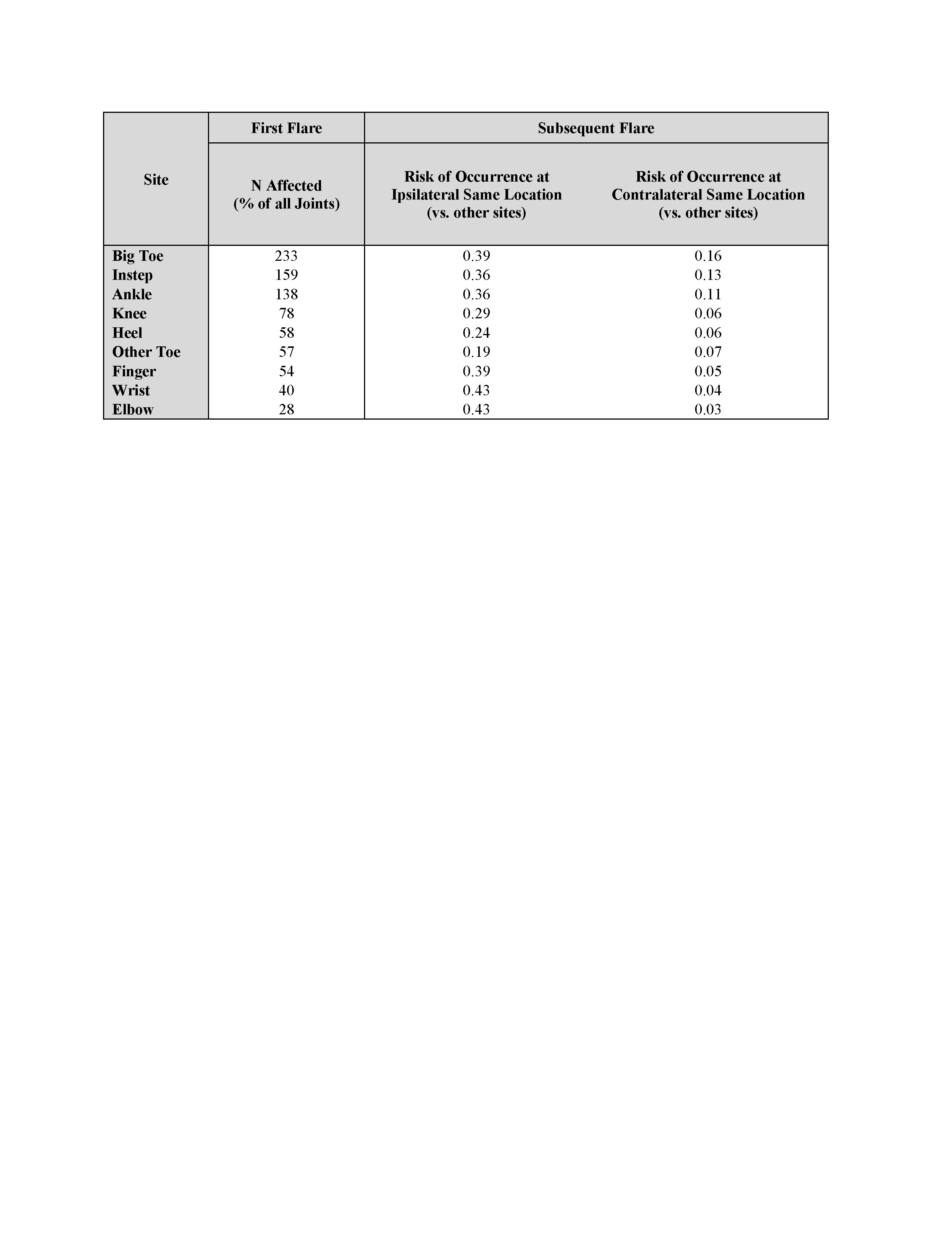 Table 1: Prevalence of Sites Affected During the First Flare, and Risk of Occurrence in Same Site During the Second Flare
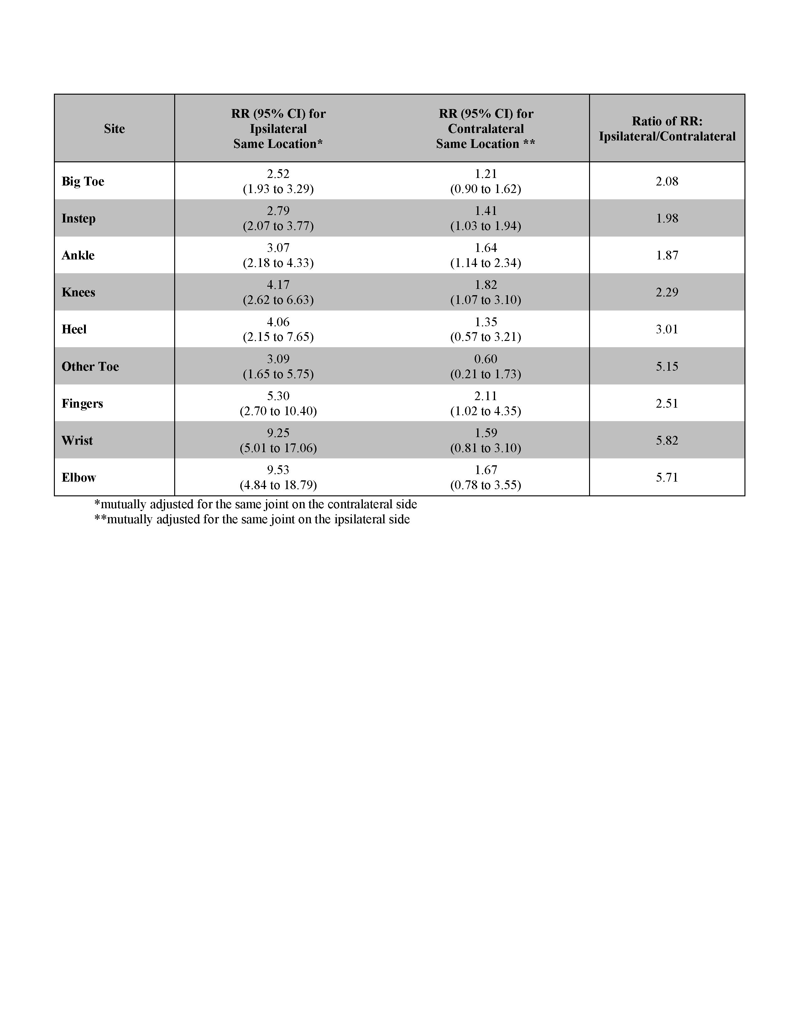 Table 2: Relative Risk (RR) of a Second Flare Occurring in the Same Site as the First Flare, on the Ipsilateral and Contralateral Sides
To cite this abstract in AMA style:
McCormick N, Yokose C, Chen C, Neogi T, Hunter D, Choi H, Zhang Y. Prospective Study of the Patterns of Joint Involvement for Sequential Gout Flares [abstract]. Arthritis Rheumatol. 2020; 72 (suppl 10). https://acrabstracts.org/abstract/prospective-study-of-the-patterns-of-joint-involvement-for-sequential-gout-flares/. Accessed .« Back to ACR Convergence 2020
ACR Meeting Abstracts - https://acrabstracts.org/abstract/prospective-study-of-the-patterns-of-joint-involvement-for-sequential-gout-flares/